
Page 73 - JSOM Summer 2025
P. 73
measurable O saturation (secondary to the severe vasocon- Patient 2’s diagnosis includes:
2
striction form hypothermia). Consider using a nasopharyn-
geal airway (NPA) or oropharyngeal airway (OPA) to assist • Accidental hypothermia II (moderate) (Table 1)
with oxygen delivery. Perform endotracheal intubation if • Aspiration pneumonitis
the equipment is available (direct or video laryngoscope). • Closed right humerus fracture
If not available, consider performing surgical cricothyrot- • Closed blunt head injury
omy. Recommend avoiding rapid sequence intubation
32
(RSI) medications, if possible, as this patient could decom- Treatment principles include the following:
pensate to the point of cardiac arrest if given strong seda-
tives and a paralytic. If patient gags or vocal cords are not • Aggressive rewarming through the use of passive external
relaxed enough to allow endotracheal tube passage, give rewarming (warm room and blankets), active external re-
RSI medications (recommend ketamine 1–2mg/kg slow warming (Bair Hugger or HPMK), and active internal warm-
IV push and rocuronium 1mg/kg IV push) with push-dose ing (warm IV fluids). Use invasive warming through the use
epinephrine readily available to supplement BP. If surgical of warm fluid bladder lavage only if absolutely necessary.
airway is performed, use 1%–2% lidocaine for local anes- • Place an internal temperature probe if available, such as
thesia to prevent the patient from reacting. a urinary catheter or nasogastric tube with built-in tem-
• Push-dose epinephrine = 1mL from 1:10,000 epinephrine sy- perature probe, or place a separate esophageal or rectal
ringe mixed with 9mL of saline from a saline flush. Concen- temperature probe. Monitor temperature continuously and
tration of epinephrine then becomes 10µg/mL, give pushes rewarm until the patient’s core temperature reaches 36°C.
of 1mL every 2–5 minutes as needed for hypotension. • As this patient has fluid aspiration into the lungs, develop-
• Place the patient on a ventilator using a lung protective ven- ment of ARDS is likely and managing the ventilator can be-
tilation strategy (6mL/kg predicted body weight). Use vol- come very difficult. Attempt maintaining O saturation of
33
2
ume assist control (V-AC) as base standard and start with 88%–95%. Use 6mL/kg of predicted body weight and slow
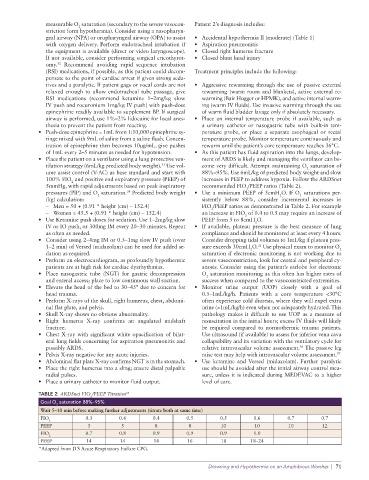
100% FIO and positive end expiratory pressure (PEEP) of increases in PEEP to address hypoxia. Follow the ARDSnet
2
5mmHg, with rapid adjustments based on peak inspiratory recommended FIO /PEEP ratios (Table 2).
2
pressures (PIP) and O saturation. Predicted body weight • Use a minimum PEEP of 5cmH O. If O saturations per-
34
2
2
2
(kg) calculation: sistently below 88%, consider incremental increases in
– Men = 50 + (0.91 * height (cm) – 152.4) FIO /PEEP ratios as demonstrated in Table 2. For example
2
– Women = 45.5 + (0.91 * height (cm) – 152.4) an increase in FIO of 0.4 to 0.5 may require an increase of
2
• Use Ketamine push doses for sedation. Use 1–2mg/kg slow PEEP from 5 to 8cmH O.
2
IV or IO push, or 300mg IM every 20–30 minutes. Repeat • If available, plateau pressure is the best measure of lung
as often as needed. compliance and should be monitored at least every 4 hours.
• Consider using 2–4mg IM or 0.5–1mg slow IV push (over Consider dropping tidal volumes to 1mL/kg if plateau pres-
35
1–2 min) of Versed (midazolam) can be used for added se- sure exceeds 30cmH O. Use physical exam to monitor O
2
2
dation as required. saturation if electronic monitoring is not working due to
• Perform an electrocardiogram, as profoundly hypothermic severe vasoconstriction, look for central and peripheral cy-
patients are at high risk for cardiac dysrhythmias. anosis. Consider using the patient’s earlobe for electronic
• Place nasogastric tube (NGT) for gastric decompression O saturation monitoring as this often has higher rates of
2
and enteral access; place to low continuous wall suction. success when compared to the vasoconstricted extremities.
• Elevate the head of the bed to 30–45° due to concern for • Monitor urine output (UOP) closely with a goal of
head trauma. 0.5–1mL/kg/h. Patients with a core temperature <30°C
• Perform X-rays of the skull, right humerus, chest, abdomi- often experience cold diuresis, where they will expel extra
nal flat plate, and pelvis. urine (>1mL/kg/h) even when not adequately hydrated. This
• Skull X-ray shows no obvious abnormality. pathology makes it difficult to use UOP as a measure of
• Right humerus X-ray confirms an angulated midshaft resuscitation in the initial hours; excess IV fluids will likely
fracture. be required compared to normothermic trauma patients.
• Chest X-ray with significant white opacification of bilat- Use ultrasound (if available) to assess for inferior vena cava
eral lung fields concerning for aspiration pneumonitis and collapsibility and its variation with the ventilatory cycle for
possibly ARDS. relative intravascular volume assessment. The passive leg
36
• Pelvis X-ray negative for any acute injuries. raise test may help with intravascular volume assessment. 37
• Abdominal flat plate X-ray confirms NGT is in the stomach. • Use ketamine and Versed (midazolam). Further paralytic
• Place the right humerus into a sling; ensure distal palpable use should be avoided after the initial airway control mea-
radial pulses. sure, unless it is indicated during MEDEVAC to a higher
• Place a urinary catheter to monitor fluid output. level of care.
TABLE 2 ARDSnet FIO /PEEP Titration 35
2
Goal O saturation 88%–95%
2
Wait 5–10 min before making further adjustments (titrate both at same time)
FIO 0.3 0.4 0.4 0.5 0.5 0.6 0.7 0.7
2
PEEP 5 5 8 8 10 10 10 12
FIO 0.7 0.8 0.9 0.9 0.9 1.0
2
PEEP 14 14 14 16 18 18–24
*Adapted from JTS Acute Respiratory Failure CPG.
Drowning and Hypothermia on an Amphibious Warship | 71