
Page 96 - JSOM Winter 2024
P. 96
Our prospective observational study sought to build upon the non-latex examination gloves. Participants were allotted a
existing body of literature related to the use of ultrasound in maximum of three minutes per tissue model; time began with
the detection of wooden FBs by describing the accuracy of placement of the ultrasound probe upon the model. They were
Special Forces (SF) medics’ ability to identify wooden FBs and asked to determine, in real time, their sonographic impres-
potentially identifying a threshold size at which a wooden FB sion of each tissue model regarding the presence or absence
can be reliably detected using ultrasound on a previously vali- of an FB. Once they had completed an assessment of one tis-
dated tissue simulation model. sue model, they were immediately moved to the next blinded
model until they had assessed all 10 models. The study PI and
AI recorded the participants impression for each model. All
Methods
study participants completed the study in one iteration and in
a single day. Pooled results of detection ability for each FB size
Study Design were reported as basic test characteristics (accuracy, sensitivity,
This study was a prospective, single-blinded, observational specificity), as well as time spent scanning each tissue model.
study conducted at the 1st Special Forces Group Conference
Room on Joint Base Lewis-McChord, WA. The Madigan Army Results
Medical Center, Department of Clinical Investigation granted
the study approval. A total of 20 medics from the 1st Special Forces Group (Air-
borne) volunteered to complete the study, performing a total
Participants of 200 scans. All participants were male, and none withdrew
A pool of 40 SF medics with no prior training in the use of from the study. Table 1 outlines sensitivity by FB size, while
POCUS for FB detection served as volunteer participant so- Table 2 outlines specificity for tissue models that did not con-
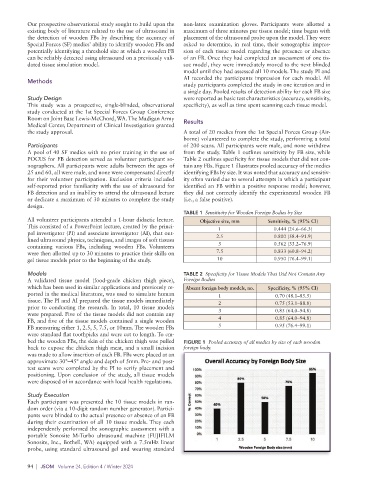
nographers. All participants were adults between the ages of tain any FBs. Figure 1 illustrates pooled accuracy of the medics
25 and 60, all were male, and none were compensated directly identifying FBs by size. It was noted that accuracy and sensitiv-
for their volunteer participation. Exclusion criteria included ity often varied due to several attempts in which a participant
self-reported prior familiarity with the use of ultrasound for identified an FB within a positive response model; however,
FB detection and an inability to attend the ultrasound lecture they did not correctly identify the experimental wooden FB
or dedicate a maximum of 30 minutes to complete the study (i.e., a false positive).
design.
TABLE 1 Sensitivity for Wooden Foreign Bodies by Size
All volunteer participants attended a 1-hour didactic lecture. Objective size, mm Sensitivity, % (95% CI)
This consisted of a PowerPoint lecture, created by the princi- 1 0.444 (24.6–66.3)
pal investigator (PI) and associate investigator (AI), that out-
lined ultrasound physics, techniques, and images of soft tissues 2.5 0.800 (58.4–91.9)
containing various FBs, including wooden FBs. Volunteers 5 0.562 (33.2–76.9)
were then allotted up to 30 minutes to practice their skills on 7.5 0.833 (60.8–94.2)
gel tissue models prior to the beginning of the study. 10 0.950 (76.4–99.1)
Models TABLE 2 Specificity for Tissue Models That Did Not Contain Any
A validated tissue model (food-grade chicken thigh piece), Foreign Bodies
which has been used in similar applications and previously re- Absent foreign body models, no. Specificity, % (95% CI)
ported in the medical literature, was used to simulate human 1 0.70 (48.1–85.5)
tissue. The PI and AI prepared the tissue models immediately 2 0.75 (53.1–88.8)
prior to conducting the research. In total, 10 tissue models
were prepared. Five of the tissue models did not contain any 3 0.85 (64.0–94.8)
FB, and five of the tissue models contained a single wooden 4 0.85 (64.0–94.8)
FB measuring either 1, 2.5, 5, 7.5, or 10mm. The wooden FBs 5 0.95 (76.4–99.1)
were standard flat toothpicks and were cut to length. To em-
bed the wooden FBs, the skin of the chicken thigh was pulled FIGURE 1 Pooled accuracy of all medics by size of each wooden
back to expose the chicken thigh meat, and a small incision foreign body.
was made to allow insertion of each FB. FBs were placed at an
approximate 30°–45° angle and depth of 5mm. Pre- and post-
test scans were completed by the PI to verify placement and
positioning. Upon conclusion of the study, all tissue models
were disposed of in accordance with local health regulations.
Study Execution
Each participant was presented the 10 tissue models in ran-
dom order (via a 10-digit random number generator). Partici-
pants were blinded to the actual presence or absence of an FB
during their examination of all 10 tissue models. They each
independently performed the sonographic assessment with a
portable Sonosite M-Turbo ultrasound machine (FUJIFILM
Sonosite, Inc., Bothell, WA) equipped with a 7.5mHz linear
probe, using standard ultrasound gel and wearing standard
94 | JSOM Volume 24, Edition 4 / Winter 2024