
Page 71 - JSOM Winter 2024
P. 71
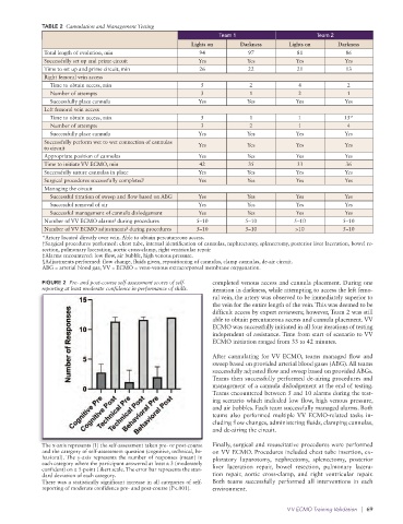
TABLE 2  Cannulation and Management Testing
                                                                   Team 1                       Team 2
                                                          Lights on      Darkness       Lights on     Darkness
              Total length of evolution, min                 94            97             81            86
              Successfully set up and prime circuit         Yes            Yes            Yes           Yes
              Time to set up and prime circuit, min          26            22             21            13
              Right femoral vein access
                Time to obtain access, min                   5              2             4              2
                Number of attempts                           3              1             2              1
                Successfully place cannula                  Yes            Yes            Yes           Yes
              Left femoral vein access
                Time to obtain access, min                   5              1             1             15*
                Number of attempts                           3              2             1              4
                Successfully place cannula                  Yes            Yes            Yes           Yes
              Successfully perform wet to wet connection of cannulas
              to circuit                                    Yes            Yes            Yes           Yes
              Appropriate position of cannulas              Yes            Yes            Yes           Yes
              Time to initiate VV ECMO, min                  42            35             33            36
              Successfully suture cannulas in place         Yes            Yes            Yes           Yes
              Surgical procedures successfully completed †  Yes            Yes            Yes           Yes
              Managing the circuit
                Successful titration of sweep and flow based on ABG  Yes   Yes            Yes           Yes
                Successful removal of air                   Yes            Yes            Yes           Yes
                Successful management of cannula dislodgement  Yes         Yes            Yes           Yes
              Number of VV ECMO alarms  during procedures   5–10          5–10           5–10           5–10
                                   ‡
              Number of VV ECMO adjustments  during procedures  5–10      5–10           >10            5–10
                                       §
              *Artery located directly over vein. Able to obtain percutaneous access.
              †Surgical procedures performed: chest tube, internal identification of cannulas, nephrectomy, splenectomy, posterior liver laceration, bowel re-
              section, pulmonary laceration, aortic cross-clamp, right ventricular repair
              ‡Alarms encountered: low flow, air bubble, high venous pressure.
              §Adjustments performed: flow change, fluids given, repositioning of cannulas, clamp cannulas, de-air circuit.
              ABG = arterial blood gas; VV = ECMO = veno-venous extracorporeal membrane oxygenation.
              FIGURE 2  Pre- and post-course self-assessment scores of self-  completed venous access and cannula placement. During one
              reporting at least moderate confidence in performance of skills.  iteration in darkness, while attempting to access the left femo-
                                                                 ral vein, the artery was observed to be immediately superior to
                                                                 the vein for the entire length of the vein. This was deemed to be
                                                                 difficult access by expert reviewers; however, Team 2 was still
                                                                 able to obtain percutaneous access and cannula placement. VV
                                                                 ECMO was successfully initiated in all four iterations of testing
                                                                 independent of assistance. Time from start of scenario to VV
                                                                 ECMO initiation ranged from 33 to 42 minutes.

                                                                 After cannulating for VV ECMO, teams managed flow and
                                                                 sweep based on provided arterial blood gases (ABG). All teams
                                                                 successfully adjusted flow and sweep based on provided ABGs.
                                                                 Teams then successfully performed de-airing procedures and
                                                                 management of a cannula dislodgement at the end of testing.
                                                                 Teams encountered between 5 and 10 alarms during the test-
                                                                 ing scenario which included low flow, high venous pressure,
                                                                 and air bubbles. Each team successfully managed alarms. Both
                                                                 teams also performed multiple VV ECMO-related tasks in-
                                                                 cluding flow changes, administering fluids, clamping cannulas,
                                                                 and de-airing the circuit.
              The x-axis represents (1) the self-assessment taken pre- or post-course   Finally, surgical and resuscitative procedures were performed
              and the category of self-assessment question (cognitive, technical, be-  on VV ECMO. Procedures included chest tube insertion, ex-
              havioral). The y-axis represents the number of responses (mean) in   ploratory laparotomy, nephrectomy, splenectomy, posterior
              each category where the participant answered at least a 3 (moderately
              confident) on a 5-point Likert scale. The error bar represents the stan-  liver laceration repair, bowel resection, pulmonary lacera-
              dard deviation of each category.                   tion repair, aortic cross-clamp, and right ventricular repair.
              There was a statistically significant increase in all categories of self-   Both teams successfully  performed all  interventions  in each
              reporting of moderate confidence pre- and post-course (P<.001).  environment.


                                                                                       V V ECMO Training Validation   |  69