
Page 69 - JSOM Winter 2024
P. 69
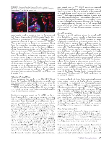
FIGURE 1  Didactic phase lighting conditions for training in    After models were on  VV ECMO, participants managed
              (A) set-up and priming of the veno-venous extracorporeal membrane     ECMO-related complications and emergencies that were ini-
              oxygenation circuit and (B) cannulation.           tiated by a reviewer in the same fashion for all iterations and
                                                                 performed damage control surgery and resuscitation proce-
                                                                 dures. 27,28  Each team performed two iterations of testing with
                                                                 white lights on and in darkness under similar conditions as di-
                                                                 dactic training. Successful completion was determined by two
                                                                 reviewers individually observing participants. A third reviewer
                                                                 was present to adjudicate divergent scores. Each reviewer had
                                                                 extensive ECMO cannulation and management experience in
                                                                 civilian hospitals. Finally, participants completed a post-course
                                                                 self-assessment.
              (A)                (B)
                                                                 Animal Preparation
              questionnaire based on questions from the  Extracorporeal   We sought to provide validation using a live animal model
              Life Support Organization (ELSO) Specialist Training Man-  given the inability to evaluate mortality and physiology using
              ual focusing on cognitive (9 questions), technical (14 ques-  a mannequin model. Practicing ECMO procedures on human
              tions), and behavioral (2 questions) aspects of VV ECMO.    volunteers is impractical and unethical; therefore, due to the
                                                            24
              The pre- and post-test was the same, and participants did not   similarities between human and swine cardiopulmonary anat-
              know the content of the knowledge assessment prior to com-  omy, we elected to use a total of 4 Yorkshire swine (Sus scrofa)
              pleting it on arrival to the course nor that they would be com-  weighing 40–70kg. These swine were fasted overnight except for
              pleting the same assessment at the end of the didactic portion   water ad lib. Swine were anesthetically induced with intramus-
              of the course. A score of 80% was considered a passing score   cular (IM) Telazol (4.4mg/kg) plus acepromazine (0.05mg/kg
              based on the development of a previous ECMO knowledge   IM). Following anesthetic induction, animal models were given
                  25
              exam.  For validation testing, several benchmarks were used   atropine sulfate (0.05mg/g IM) and buprenorphine (0.02mg/kg
              to measure success in performing tasks in a time-sensitive   IM), and endotracheal intubation was performed. Isoflurane
              manner. Previous studies have demonstrated that VV ECMO   anesthesia was delivered using the A.D.S 2000 Veterinary An-
              cannulation can take between 45 to 60 minutes from the start   esthesia Delivery System a microprocessor controlled anes-
              of the procedure to initiating VV ECMO.  Time to circuit   thetic ventilator. Following induction of anesthesia, vital signs
                                                26
              priming and set-up takes approximately 20 to 30 minutes,   were continuously monitored using a Surgivet Advisor Tech
              according to unpolished institutional data of novice trainees.   monitor with 3-lead ECG, standard SPO , end-tidal CO , and
                                                                                                2
                                                                                                            2
              Demographic information was collected at the beginning of   rectal temperature probe. Thermal support was provided us-
              the course. Total instructional time at STC was 17 hours over   ing a 3M Bair Hugger Warming Blanket System and isotonic
              2 training days.                                   intravenous (IV) fluid support (0.9% NaCl) was delivered via
                                                                 an ear vein IV catheter.
              Validation (Testing) Phase
              Course participants then traveled to the 81st MDG CRL at   To prevent cardiac dysrhythmias during guidewire placement,
              Keesler Air Force Base (AFB). Participants were divided into   animal models were given a 2–3mg/kg lidocaine bolus prior
              two teams and tested on their ability to set up and prime the   to cannulation and maintained on a 50µg/kg/min lidocaine
              VV ECMO circuit and place Yorkshire swine (Sus scrofa) on   constant-rate Infusion (CRI) until cannulation was complete.
              VV ECMO using the provided checklists. A 2-hour timeframe   Following  confirmation  of  successful  VV  ECMO  initiation
              was allotted per testing event. Testing was marked complete   and swine stabilization, animal models were given a 100mg
              when all objectives were complete.                 bolus of ketamine and switched from gas anesthesia to total
                                                                 intravenous anesthesia (TIVA) CRI (ketamine 12–48mg/kg/hr
              Participants cannulated models for  VV ECMO via the bi-  and midazolam 0.5–1.5mg/kg/hr). Prior thoracotomy proce-
              femoral  approach. Two  surgeons,  one  emergency  physician,   dures animal models were transitioned back to gas anesthesia.
              and one anesthesiologist performed one access, dilation, and   Upon completion of all VV ECMO training procedures animal
              cannulation. Teams determined who would perform the ac-  models were euthanized with an overdose (1mL/10 pounds)
              cess and cannulations based on who would be most likely to   of Euthasol (sodium pentobarbital 390mg/mL and phenytoin
              perform these skills in the operational environment. Cannu-  50mg/mL) given intravenously or via intracardiac injection.
              lation was accomplished using ultrasound-guided access and
              percutaneous Seldinger technique. Participants had the option   Data Storage and Analysis
              of using a 5-French micropuncture kit (G48007, Cook Med-  All participants were volunteers who signed consent to partic-
              ical, Bloomington, IN) or using a needle in the dilator kit. A   ipate in research prior to the course start. Study data were col-
              guidewire was placed and serial dilations up to a 16-French   lected and managed using Research Electronic Data Capture
              dilator of the femoral veins were performed using a percuta-  (REDCap) tools hosted at our institution. 29,30  Demographics,
              neous insertion kit (LivaNova Sorin 8Fr-24Fr REF: 200-120).   pre- and post-course self-assessments and pre- and post-course
              The 19-French return cannula was placed in the right femoral   knowledge assessments were collected from participants.
              vein and the 19-French drainage cannula was placed in the left
              femoral vein. The Maquet PVS 1938 cannula was used as the   Data were analyzed with descriptive statistics. Parametric or
              drainage cannula and the Medtronic Bio-Medicus 96670-019   nonparametric statistics were used based on the nature of the
              was used as the return cannula. Cannulas were de-aired and   data. Normality was assessed with the Shapiro-Wilk test
              attached to the ECMO circuit through a wet-to-wet connec-  and examination  of stem-and-leaf  as well  as  q-q plots.  All
              tion. The cannulas were then sutured in place for stabilization.  data were normally distributed and presented as means and

                                                                                       V V ECMO Training Validation   |  67