
Page 78 - JSOM Summer 2023
P. 78
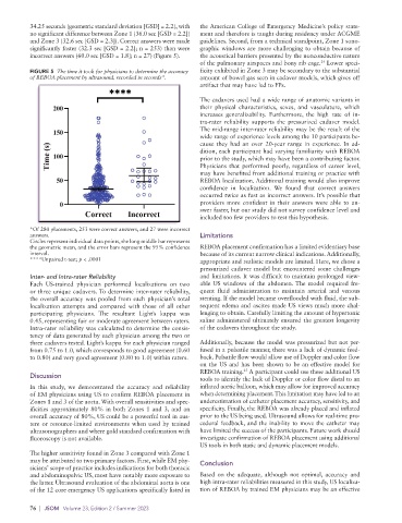
34.25 seconds (geometric standard deviation [GSD] = 2.2), with   the American College of Emergency Medicine’s policy state-
          no significant difference between Zone 1 (36.0 sec [GSD = 2.2])   ment and therefore is taught during residency under ACGME
          and Zone 3 (32.6 sec [GSD = 2.3]). Correct answers were made   guidelines. Second, from a technical standpoint, Zone 1 sono-
          significantly faster (32.3 sec [GSD = 2.2]; n = 253) than were   graphic windows are more challenging to obtain because of
          incorrect answers (60.0 sec [GSD = 1.8]; n = 27) (Figure 5).  the acoustical barriers presented by the nonconductive nature
                                                             of the pulmonary airspaces and bony rib cage.  Lower speci-
                                                                                                 24
          FIGURE 5  The time it took for physicians to determine the accuracy   ficity exhibited in Zone 3 may be secondary to the substantial
          of REBOA placement by ultrasound, recorded in seconds*.  amount of bowel gas seen in cadaver models, which gives off
                                                             artifact that may have led to FPs.

                                                             The cadavers used had a wide range of anatomic variants in
                                                             their physical characteristics, sexes, and vasculature, which
                                                             increases generalizability. Furthermore, the high rate of in-
                                                             tra-rater reliability supports the pressurized cadaver model.
                                                             The mid-range inter-rater reliability may be the result of the
                                                             wide range of experience levels among the 10 participants be-
                                                             cause they had an over 20-year range in experience. In ad-
                                                             dition, each participant had varying familiarity with REBOA
                                                             prior to the study, which may have been a contributing factor.
                                                             Physicians that performed poorly, regardless of career level,
                                                             may have benefited from additional training or practice with
                                                             REBOA localization. Additional training would also improve
                                                             confidence  in  localization.  We  found that  correct  answers
                                                             occurred twice as fast as incorrect answers. It’s possible that
                                                             providers more confident in their answers were able to an-
                                                             swer faster, but our study did not survey confidence level and
                                                             included too few providers to test this hypothesis.
          *Of 280 placements, 253 were correct answers, and 27 were incorrect
          answers.                                           Limitations
          Circles represent individual data points, the long middle bar represents
          the geometric mean, and the error bars represent the 95% confidence   REBOA placement confirmation has a limited evidentiary base
          interval.                                          because of its current narrow clinical indications. Additionally,
          ****Unpaired t-test; p < .0001                     appropriate and realistic models are limited. Here, we chose a
                                                             pressurized cadaver model but encountered some challenges
          Inter- and Intra-rater Reliability                 and limitations. It was difficult to maintain prolonged view-
          Each US-trained physician performed localizations on two   able US windows of the abdomen. The model required fre-
          or three unique cadavers. To determine inter-rater reliability,   quent  fluid administration to  maintain  arterial  and venous
          the overall accuracy was pooled from each physician’s total   stenting. If the model became overflooded with fluid, the sub-
          localization attempts and compared with those of all other   sequent edema and ascites made US views much more chal-
          participating physicians.  The resultant Light’s kappa was   lenging to obtain. Carefully limiting the amount of hypertonic
          0.45, representing fair or moderate agreement between raters.   saline administered ultimately ensured the greatest longevity
            Intra-rater reliability was calculated to determine the consis-  of the cadavers throughout the study.
          tency of data generated by each physician among the two or
          three cadavers tested. Light’s kappa for each physician ranged   Additionally, because the model was pressurized but not per-
          from 0.75 to 1.0, which corresponds to good agreement (0.60   fused in a pulsatile manner, there was a lack of dynamic feed-
          to 0.80) and very good agreement (0.80 to 1.0) within raters.  back. Pulsatile flow would allow use of Doppler and color flow
                                                             on the US and has been shown to be an effective model for
                                                                          25
                                                             REBOA training.  A participant could use these additional US
          Discussion                                         tools to identify the lack of Doppler or color flow distal to an
          In  this  study,  we  demonstrated  the  accuracy  and  reliability   inflated aortic balloon, which may allow for improved accuracy
          of EM physicians using US to confirm REBOA placement in   when determining placement. This limitation may have led to an
          Zones 1 and 3 of the aorta. With overall sensitivities and spec-  underestimation of catheter placement accuracy, sensitivity, and
          ificities approximately 80% in both Zones 1 and 3, and an   specificity. Finally, the REBOA was already placed and inflated
          overall accuracy of 80%, US could be a powerful tool in aus-  prior to the US being used. Ultrasound allows for real-time pro-
          tere or resource-limited environments when used by trained   cedural feedback, and the inability to move the catheter may
          ultrasonographers and where gold standard confirmation with   have limited the success of the participants. Future work should
          fluoroscopy is not available.                      investigate confirmation of REBOA placement using additional
                                                             US tools in both static and dynamic placement models.
          The higher sensitivity found in Zone 3 compared with Zone 1
          may be attributed to two primary factors. First, while EM phy-  Conclusion
          sicians’ scope of practice includes indications for both thoracic
          and abdominopelvic US, most have notably more exposure to   Based on the adequate, although not optimal, accuracy and
          the latter. Ultrasound evaluation of the abdominal aorta is one   high intra-rater reliabilities measured in this study, US localiza-
          of the 12 core emergency US applications specifically listed in   tion of REBOA by trained EM physicians may be an effective

          76  |  JSOM   Volume 23, Edition 2 / Summer 2023