
Page 75 - JSOM Fall 2022
P. 75
9
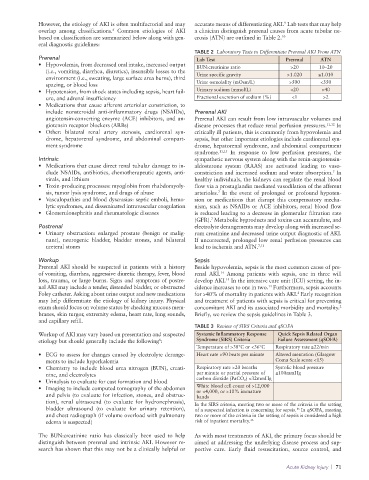
However, the etiology of AKI is often multifactorial and may accurate means of differentiating AKI. Lab tests that may help
6
overlap among classifications. Common etiologies of AKI a clinician distinguish prerenal causes from acute tubular ne
based on classification are summarized below along with gen crosis (ATN) are outlined in Table 2. 10
eral diagnostic guidelines:
TABLE 2 Laboratory Tests to Differentiate Prerenal AKI From ATN
Prerenal Lab Test Prerenal ATN
• Hypovolemia, from decreased oral intake, increased output BUN:creatinine ratio >20 10–20
(i.e., vomiting, diarrhea, diuretics), insensible losses to the
environment (i.e., sweating, large surface area burns), third Urine specific gravity >1.020 ≤1.010
spacing, or blood loss Urine osmolality (mOsm/L) >500 <350
• Hypotension, from shock states including sepsis, heart fail Urinary sodium (mmol/L) <20 >40
ure, and adrenal insufficiency Fractional excretion of sodium (%) <1 >2
• Medications that cause afferent arteriolar constriction, to
include nonsteroidal antiinflammatory drugs (NSAIDs), Prerenal AKI
angiotensinconverting enzyme (ACE) inhibitors, and an Prerenal AKI can result from low intravascular volumes and
giotensin receptor blockers (ARBs) disease processes that reduce renal perfusion pressures. 11,12 In
• Other: bilateral renal artery stenosis, cardiorenal syn critically ill patients, this is commonly from hypovolemia and
drome, hepatorenal syndrome, and abdominal compart sepsis, but other important etiologies include cardiorenal syn
ment syndrome drome, hepatorenal syndrome, and abdominal compartment
syndrome. 11,13 In response to low perfusion pressures, the
Intrinsic sympathetic nervous system along with the renin angiotensin
• Medications that cause direct renal tubular damage to in aldosterone system (RAAS) are activated leading to vaso
clude NSAIDs, antibiotics, chemotherapeutic agents, anti constriction and increased sodium and water absorption. In
7
virals, and lithium healthy individuals, the kidneys can regulate the renal blood
• Toxinproducing processes: myoglobin from rhabdomyoly flow via a prostaglandin mediated vasodilation of the afferent
sis, tumor lysis syndrome, and drugs of abuse arterioles. In the event of prolonged or profound hypoten
7
• Vasculopathies and blood dyscrasias: septic emboli, hemo sion or medications that disrupt this compensatory mecha
lytic syndromes, and disseminated intravascular coagulation nism, such as NSAIDs or ACE inhibitors, renal blood flow
• Glomerulonephritis and rheumatologic diseases is reduced leading to a decrease in glomerular filtration rate
(GFR). Metabolic byproducts and toxins can accumulate, and
7
Postrenal electrolyte derangements may develop along with increased se
• Urinary obstruction: enlarged prostate (benign or malig rum creatinine and decreased urine output diagnostic of AKI.
nant), neurogenic bladder, bladder stones, and bilateral If uncorrected, prolonged low renal perfusion pressures can
ureteral stones lead to ischemia and ATN. 7,12
Workup Sepsis
Prerenal AKI should be suspected in patients with a history Beside hypovolemia, sepsis is the most common cause of pre
of vomiting, diarrhea, aggressive diuretic therapy, fever, blood renal AKI. Among patients with sepsis, one in three will
14
loss, trauma, or large burns. Signs and symptoms of postre develop AKI. In the intensive care unit (ICU) setting, the in
14
nal AKI may include a tender, distended bladder, or obstructed cidence increases to one in two. Furthermore, sepsis accounts
14
Foley catheter. Asking about urine output and new medications for >40% of mortality in patients with AKI. Early recognition
8
may help differentiate the etiology of kidney injury. Physical and treatment of patients with sepsis is critical for preventing
exam should focus on volume status by checking mucous mem concomitant AKI and its associated morbidity and mortality.
2
branes, skin turgor, extremity edema, heart rate, lung sounds, Briefly, we review the sepsis guidelines in Table 3.
and capillary refill.
TABLE 3 Review of SIRS Criteria and qSOFA
Workup of AKI may vary based on presentation and suspected Systemic Inflammatory Response Quick Sepsis Related Organ
8
etiology but should generally include the following : Syndrome (SIRS) Criteria Failure Assessment (qSOFA)
Temperature of >38°C or <36°C Respiratory rate ≥22/min
• ECG to assess for changes caused by electrolyte derange Heart rate >90 beats per minute Altered mentation (Glasgow
ments to include hyperkalemia Coma Scale score <15)
• Chemistry to include blood urea nitrogen (BUN), creati Respiratory rate >20 breaths Systolic blood pressure
nine, and electrolytes per minute or partial pressure of ≤100mmHg
• Urinalysis to evaluate for cast formation and blood carbon dioxide (PaCO 2 ) <32mmHg
• Imaging to include computed tomography of the abdomen White blood cell count of >12,000
or <4,000, or >10% immature
and pelvis (to evaluate for infection, stones, and obstruc bands
tion), renal ultrasound (to evaluate for hydronephrosis), In the SIRS criteria, meeting two or more of the criteria in the setting
bladder ultrasound (to evaluate for urinary retention), of a suspected infection is concerning for sepsis. In qSOFA, meeting
15
and chest radiograph (if volume overload with pulmonary two or more of the criteria in the setting of sepsis is considered a high
edema is suspected) risk of inpatient mortality. 16
The BUN:creatinine ratio has classically been used to help As with most treatments of AKI, the primary focus should be
distinguish between prerenal and intrinsic AKI. However re aimed at addressing the underlying disease process and sup
search has shown that this may not be a clinically helpful or portive care. Early fluid resuscitation, source control, and
Acute Kidney Injury | 71