
Page 72 - JSOM Summer 2022
P. 72
overall oxygen-carrying capacity of RBCs. Hereditary factors some settings the diagnosis may be found incidentally. An ef-
include methemoglobinemia and the hemoglobin S variant, fort should be made to correlate low hemoglobin values with
the latter of which is present in sickle cell disease. Hemoglobin patient history, physical examination, and other laboratory
structural alterations underlie these pathologies, referred to as findings, when available, to identify an underlying cause. Al-
hemoglobinopathies, and affect not only the oxygen-carrying though laboratory values such as mean corpuscular volume
capacity but also RBC lifespan and tendency toward destruc- (MCV) and RBC distribution width (RDW) can narrow pos-
tion (i.e., hemolysis). Environmental factors may likewise sible causes of anemia, these may be difficult to obtain in the
worsen hereditary disease or alone cause hemoglobinopathy. austere setting. 1–3,9,20
Examples include sulfhemoglobinemia (i.e., exposure to sulfa),
cyanohemoglobinemia (i.e., exposure to cyanide), and carbon
monoxide toxicity. 1–3,20,23 Hemoglobinopathy can compromise Patient Presentation and Assessment
tissue oxygenation and trigger compensatory responses, such Initial assessment in suspected or confirmed cases of anemia
1,9
as increased cardiac output and vasoconstriction. A right- immediately focuses on hemodynamic stability and the search
ward shift in the oxyhemoglobin dissociation curve increases for possible sources of gross hemorrhage. Priority should be
oxygen release from circulating hemoglobin to supplement made for stabilization in all patients, with or without signifi-
tissue oxygenation. 9,22 If anemia continues over several weeks cant hemorrhage. 1–3,26,27 It is important to note, however, that
or longer, hematologic adaptations occur, such as increasing RBCs can carry up to four times the oxygen required for the
plasma volume to offset lost intravascular mass and eryth- body at rest and therefore can compensate for hypoxemia,
ropoietin surges to promote RBC production. 1,9,22 Increased precluding tachypnea or tachycardia. 1,22,26 Stable vital signs
RBC turnover and a hastened production cycle increase the should not necessarily reassure medics. 1,22,26 When vital signs
proportion of circulating immature reticulocytes. 1,18,22,24 These are unstable in suspected anemia, medics should refrain from
physiologic adaptations are important to remember during reflexive fluid resuscitation with crystalloids or other non-
the initial evaluation because they can prevent compensatory blood products because military and civilian trauma literature
physiologic responses, concealing outward signs and symp- continues to illustrate the harmful effect of dilution. 28–30
toms classically associated with anemia. 2,3,6,22
In the anemic patient, medics should consider all possible
sources of recent or ongoing hemorrhage, including the nose
Classification
(epistaxis), lungs (hemoptysis), gastrointestinal tract (hemate-
Evaluation of anemia in the forward-deployed setting neces- mesis, hematochezia, melena), and genitourinary (hematuria)
sitates a foundational understanding of acute and chronic systems, whereas others may be based on patient demograph-
causes in relation to overall patient stability. 1–3,24 Ranges for ics, such as ectopic pregnancy in women of childbearing
acute and chronic, often used to describe medical conditions, age. 1,3,31 A thorough surgical and medical history may suggest
are not equally well-defined in anemia but rather are broadly other conditions indicating proneness to bleeding, including
used in patient evaluations. peptic ulcer disease, Crohn’s disease, and ulcerative colitis.
32
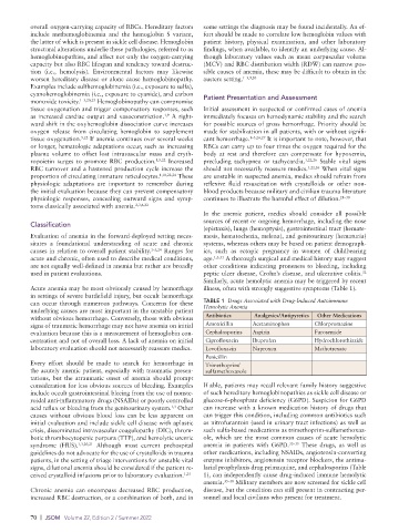
Similarly, acute hemolytic anemia may be triggered by recent
Acute anemia may be most obviously caused by hemorrhage illness, often with strongly suggestive symptoms (Table 1).
in settings of severe battlefield injury, but occult hemorrhage
can occur through numerous pathways. Concerns for these TABLE 1 Drugs Associated with Drug-Induced Autoimmune
underlying causes are most important in the unstable patient Hemolytic Anemia
without obvious hemorrhage. Conversely, those with obvious Antibiotics Analgesics/Antipyretics Other Medications
signs of traumatic hemorrhage may not have anemia on initial Amoxicillin Acetaminophen Chlorpromazine
evaluation because this is a measurement of hemoglobin con- Cephalosporins Aspirin Furosemide
centration and not of overall loss. A lack of anemia on initial Ciprofloxacin Ibuprofen Hydrochlorothiazide
laboratory evaluation should not necessarily reassure medics. Levofloxacin Naproxen Methotrexate
Penicillin
Every effort should be made to search for hemorrhage in Trimethoprim/
the acutely anemic patient, especially with traumatic presen- sulfamethoxazole
tations, but the atraumatic onset of anemia should prompt
consideration for less obvious sources of bleeding. Examples If able, patients may recall relevant family history suggestive
include occult gastrointestinal bleeing from the use of nonste- of such hereditary hemoglobinopathies as sickle cell disease or
roidal anti-inflammatory drugs (NSAIDs) or poorly controlled glucose-6-phosphate deficiency (G6PD). Suspicion for G6PD
acid reflux or bleeding from the genitourinary system. Other can increase with a known medication history of drugs that
1,3
causes without obvious blood loss can be less apparent on can trigger this condition, including common antibiotics such
initial evaluation and include sickle cell disease with aplastic as nitrofurantoin (used in urinary tract infections) as well as
crisis, disseminated intravascular coagulopathy (DIC), throm- such sulfa-based medications as trimethoprim-sulfamethoxaz-
botic thrombocytopenic purpura (TTP), and hemolytic uremic ole, which are the most common causes of acute hemolytic
syndrome (HUS). 1,3,20,21 Although most current prehospital anemia in patients with G6PD. 33–35 These drugs, as well as
guidelines do not advocate for the use of crystalloids in trauma other medications, including NSAIDs, angiotensin-converting
patients, in the setting of triage interventions for unstable vital enzyme inhibitors, angiotensin receptor blockers, the antima-
signs, dilutional anemia should be considered if the patient re- larial prophylaxis drug primaquine, and cephalosporins (Table
ceived crystalloid infusions prior to laboratory evaluation. 1,25 1), can independently cause drug-induced immune hemolytic
anemia. 35–38 Military members are now screened for sickle cell
Chronic anemia can encompass decreased RBC production, disease, but the condition can still present in contracting per-
increased RBC destruction, or a combination of both, and in sonnel and local civilians who present for treatment.
70 | JSOM Volume 22, Edition 2 / Summer 2022