
Page 66 - JSOM Summer 2022
P. 66
Elevated serum lactate, the direct metabolic product of an- parasympathetic stimulation. Anaphylactic shock is also dis-
aerobic metabolism, is an important surrogate for organ dys- tributive in nature. It refers to severe and life- threatening sys-
2
function and is an independent marker for sepsis severity. temic reactions commonly involving IgE-dependent pathways.
Leukocytosis with or without bandemia, blood sugar derange- Anaphylactic shock can also involve a nonimmunologic direct
ments, elevated serum creatinine, thrombocytopenia, and hy- release of histamine and other mediators from mast cells and
perbilirubinemia are common laboratory signs in shock across basophils; this has been previously referred to as the “anaphy-
etiologies. lactoid reaction.”
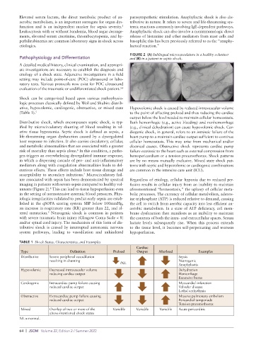
FIGURE 2 (A) Sublingual microvasculature in a healthy volunteer
Pathophysiology and Differentiation and (B) in a patient in septic shock.
A detailed medical history, clinical examination, and appropri-
ate investigations are necessary to establish the diagnosis and
etiology of a shock state. Adjunctive investigations in a field
setting may include point-of-care (POC) ultrasound or labo-
ratory tests. Various protocols now exist for POC ultrasound
evaluation of the traumatic or undifferentiated shock patient. 3,4
Shock can be categorized based upon various pathophysio-
logic processes classically defined by Weil and Shubin: distrib-
utive, hypovolemic, cardiogenic, obstructive, or mixed state Hypovolemic shock is caused by reduced intravascular volume
(Table 1). 5 to the point of affecting preload and thus reducing the cardiac
output below the level needed to maintain cellular homeostasis.
Distributive shock, which encompasses septic shock, is typ- Both hemorrhagic (e.g., active bleeding) and nonhemorrhage
ified by microcirculatory shunting of blood resulting in rel- (e.g., clinical dehydration) can cause hypovolemic shock. Car-
ative tissue hypoxemia. Septic shock is defined as sepsis, a diogenic shock, in general, refers to an intrinsic failure of the
life-threatening organ dysfunction caused by a dysregulated heart pump to a maintain cardiac output sufficient to continue
host response to infection. It also carries circulatory, cellular, cellular homeostasis. This may arise from mechanical and/or
and metabolic abnormalities that are associated with a greater electrical causes. Obstructive shock represents cardiac pump
risk of mortality than sepsis alone. In this condition, a patho- failure extrinsic to the heart such as external compression from
6
gen triggers an overwhelming dysregulated immune response, hemopericardium or a tension pneumothorax. Shock patterns
in which a dispersing cascade of pro- and anti- inflammatory are by no means mutually exclusive. Mixed state shock pat-
mediators along with coagulation abnormalities leads to del- terns with septic and hypovolemic or cardiogenic combinations
eterious effects. These effects include host tissue damage and are common in the intensive care unit (ICU).
susceptibility to secondary infections. Microcirculatory fail-
7
ure associated with sepsis has been demonstrated by spectral Regardless of etiology, cellular hypoxia due to reduced per-
imaging in patients with severe sepsis compared to healthy vol- fusion results in cellular injury from an inability to maintain
8
unteers (Figure 2). This can lead to tissue hypoperfusion even aforementioned “homeostasis,” the upkeep of cellular meta-
in the setting of normotensive systemic blood pressures. Phys- bolic processes. The currency of cellular metabolism, adenos-
iologic irregularities validated to predict early sepsis are estab- ine triphosphate (ATP) is reduced relative to demand, causing
lished in the qSOFA scoring system: SBP below 100mmHg, the cell to switch from aerobic capacity into less efficient an-
an increase in respiratory rate (RR) greater than 22, and al- aerobic metabolism. In a state of ATP deficiency, cell mem-
9
tered mentation. Neurogenic shock is common in patients brane dysfunction then manifests as an inability to maintain
with severe traumatic brain injury (Glasgow Coma Scale < 8) the contents of both the intra- and extracellular spaces. Serum
and/or spinal cord injury. The mechanism of this form of dis- lactate levels subsequently rise. When this process extends
tributive shock is caused by interrupted autonomic nervous to the tissue level, it becomes self-perpetuating and worsens
system pathways, leading to vasodilation and unhindered hypoperfusion.
TABLE 1 Shock States, Characteristics, and Examples
Cardiac
Type Definition Preload Output Afterload Examples
Distributive Severe peripheral vasodilation Sepsis
resulting in shunting Neurogenic
/NL Anaphylactic
Hypovolemic Decreased intravascular volume Dehydration
reducing cardiac output Hemorrhage
Extensive burns
Cardiogenic Intracardiac pump failure causing Myocardial infarction
reduced cardiac output Valvular disease
Lethal arrhythmia
Obstructive Extracardiac pump failure causing Massive pulmonary embolism
reduced cardiac output Pericardial tamponade
Tension pneumothorax
Mixed Overlap of two or more of the Variable Variable Variable Acute pericarditis
above-mentioned shock states
NL = normal.
64 | JSOM Volume 22, Edition 2 / Summer 2022