
Page 46 - JSOM Summer 2022
P. 46
Myoglobin, a protein that carries oxygen within the muscle, FIGURE 2 Complications of crush injury.
damages the renal system by precipitating rhabdomyolysis
when circulating at high levels. Crush injuries create an enor-
mous amount of circulating myoglobin, which is not normally
found in blood. Normally, the glomeruli in the kidneys filter
myoglobin, but once renal threshold is surpassed, the convo-
luted distal tubules become obstructed, which leads to kidney
dysfunction. Additionally, the increasing collection of myoglo-
bin creates localized vasoconstriction, further exacerbating the
problem of filtering out toxic metabolites.
Acute (traumatic) rhabdomyolysis caused by a rapid release
of myoglobin may be more damaging than extended ischemia
of soft tissue. During reperfusion, reactive hyperemia and in-
creased capillary permeability causes intravascular hypovole-
mia and localized tissue edema, further increasing the potential
for compartment syndrome as more fluid builds in the muscle and amount (COCA) of the patient’s urine. A patient expe-
contained by inflexible fascia. riencing rhabdomyolysis will have tea or cola-colored urine.
As a mnemonic, the Special Operations medic can remember
The release of ions by damaged skeletal muscle cells affects the “COCA-cola equals rhabdomyolysis.” This alone can indicate
surrounding cells by changing the ratio of intracellular and ex- muscle cell breakdown or kidney dysfunction without the ne-
tracellular components. Cells will third space intracellular fluid cessity of carrying an extra diagnostic tool. Utilizing a urine
through osmosis in an effort to solve the problem. The result is dipstick is better, as detecting and trending myoglobinuria is
damaged yet intact cells that initiate an inflammatory cascade, another means to identify the level of severity of muscle tissue
sending cytokines and proinflammatory markers to the injury breakdown. Urine dipsticks can detect myoglobin but will also
site. Cytokines and inflammatory markers exacerbate local in- present positive for hemoglobin, leading to a lack of speci-
flammation and the potential for compartment syndrome. ficity. However, in a resource-strained environment, this tool
can still be useful, especially if there is an extended evacuation
As the muscle cells increase in size due to the compression on time. Best practice would be analyzing serum levels of myoglo-
the limb and inflammatory response, the muscle tissue becomes bin, requiring laboratory support. For a SOF team, this may be
compressed within the fascia, creating a host of new problems. achievable in some environments due to the nature of partner-
The fascia normally serves as a durable connective tissue en- ships built with governments and local civilian organizations.
closure for the muscle compartments, within which the muscle
fibers can contract. As pressure rises within this enclosure, how- Any means to measure the patient’s urine output is necessary
ever, the perfusion of the capillaries, nerves, and cells can become to gain a better picture of their clinical presentation. A medic
compromised and can result in ischemic destruction to these tis- needs to have a way to catch the patient’s urine, utilizing a wa-
sues. Tissue edema and subsequent swelling maximizes in 1–2 ter bottle at a minimum and estimating urine output. A better
days; however, postinjury reperfusion can appear anywhere be- solution is to catch urine in a water bottle and use a syringe to
2
tween 2 and 5 days. Compartment pressures above 30mmHg determine the patient’s exact urine output. Best practice would
sustained for 6–8 hours can lead to irreversible tissue damage. 4 be to place a transurethral indwelling “Foley” catheter, as it is
the preferred way to monitor urine output. The goal of urine
Additionally, occlusion of vessels by traumatic compression of output is 100–200mL/hr. 5
soft tissue prevents oxygen from reaching the limb and the
removal of toxins and damaged cells, leading to ischemia and Cardiac monitoring is paramount postextrication due to the
localized edema. Cells not receiving oxygen must revert to an- influx of intracellular potassium and calcium released system-
aerobic metabolism to conduct respiration, creating lactic acid ically. At minimum, close monitoring of vital signs and cir-
as a byproduct. The buildup of lactic acid decreases tissue pH, culation, examination for premature ventricular contractions
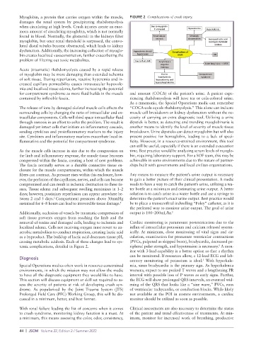
causing metabolic acidosis. Each of these changes lead to sys- (PVCs, palpated as skipped beats), bradycardia, decreased pe-
5
temic complications, detailed in Figure 2. ripheral pulse strength, and hypotension is necessary. A mon-
itor with 3-lead capability is a better option so that a rhythm
can be monitored. If resources allow, a 12-lead ECG and lab-
Diagnosis
oratory monitoring of potassium is ideal. With hyperkale-
5
Special Operations medics often work in resource-constrained mia, sinus bradycardia is the primary sign. As hyperkalemia
environments, in which the mission may not allow the medic worsens, expect to see peaked T waves and a lengthening PR
to have all the diagnostic equipment they would like to have. interval with possible loss of P waves as early signs. Further,
This section will discuss equipment or skill set required to as- the ECG will show prolonged QRS intervals, an eventual wid-
sess the severity of patients at risk of developing crush syn- ening of the QRS that looks like a “sine wave,” PVCs, runs
drome. As popularized by the Joint Trauma System (JTS) of ventricular tachycardia, or conduction blocks. While likely
Prolonged Field Care (PFC) Working Group, this will be dis- not available at the POI in austere environments, a cardiac
cussed in a minimum, better, and best format. monitor should be utilized as soon as possible.
With renal failure leading the list of concerns when it comes Clinical assessments are also necessary to determine the status
to crush syndrome, monitoring kidney function is a must. At of the patient and trend effectiveness of treatments. At min-
a minimum, this means assessing the color, odor, consistency, imum, monitor for increased work of breathing, productive
44 | JSOM Volume 22, Edition 2 / Summer 2022