
Page 47 - JSOM Spring 2021
P. 47
Methods at a sample rate of 4 Hz. Each device was tightened as much as
possible. Four researchers shared the responsibility of applying
Commercially purchased force-sensitive resistors (FSRs) (FSR the five pelvic compression devices to each subject.
Model 406; Interlink Electronics; Food and Drug Administra-
tion approved) were calibrated and used for force assessments Descriptions of Compression Devices
(Figure 1A). In a closed circuit, the application of force to an and Their Applications
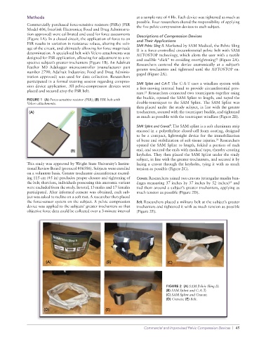
FSR results in variation in resistance values, altering the volt- SAM Pelvic Sling II. Marketed by SAM Medical, the Pelvic Sling
age of the circuit, and ultimately allowing for force magnitude II is a force-controlled circumferential pelvic belt with SAM
determination. A specialized belt with Velcro attachments was AUTOSTOP technology, which alerts the user with a tactile
designed for FSR application, allowing for adjustment to a re- and audible “click” to avoiding overtightening (Figure 2A).
22
spective subject’s greater trochanters (Figure 1B). An Adafruit Researchers centered the device anatomically at a subject’s
Feather M0 Adalogger microcontroller (manufacturer part greater trochanters and tightened until the AUTOSTOP en-
number 2798; Adafruit Industries; Food and Drug Adminis- gaged (Figure 2A).
tration approved) was used for data collection. Researchers
participated in a formal training session regarding compres- SAM Splint and C-A-T. The C-A-T uses a windlass system with
sion device application. All pelvic-compression devices were a free-moving internal band to provide circumferential pres-
placed and secured atop the FSR belt.
sure. Researchers connected two tourniquets together using
23
the buckle, opened the SAM Splint to length, and taped the
FIGURE 1 (A) Force-sensitive resistor (FSR); (B) FSR belt with
Velcro attachments. double-tourniquet to the SAM Splint. The SAM Splint was
then placed under the study subject, in line with the greater
(A) trochanters, secured with the tourniquet buckle, and tightened
as much as possible with the tourniquet windlass (Figure 2B).
SAM Splint and Cravat . The SAM splint is a soft aluminum strip
®
encased in a polyethylene closed-cell foam coating, designed
to be a compact, lightweight device for the immobilization
of bone and stabilization of soft-tissue injuries. Researchers
24
opened the SAM Splint to length, folded a portion of each
(B) end, and secured the ends with medical tape, thereby creating
keyholes. They then placed the SAM Splint under the study
subject, in line with the greater trochanters, and secured it by
This study was approved by Wright State University’s Institu- lacing a cravat through the keyholes, tying it with as much
tional Review Board (protocol #06586). Subjects were enrolled tension as possible (Figure 2C).
on a volunteer basis. Greater trochanter circumference exceed-
ing 115 cm (45 in) precludes proper closure and tightening of Cravats. Researchers joined two cravats (triangular muslin ban-
the belt; therefore, individuals possessing this anatomic variant dages measuring 37 inches by 37 inches by 52 inches) and
25
were excluded from the study. In total, 13 males and 17 females tied them around a subject’s greater trochanters, applying as
participated. After informed consent was obtained, each sub- much tension as possible (Figure 2D).
ject was asked to recline on a soft mat. A researcher then placed
the force-sensor system on the subject. A pelvic compression Belt. Researchers placed a military belt at the subject’s greater
device was applied to the subjects’ greater trochanters so that trochanters and tightened it with as much tension as possible
objective force data could be collected over a 3-minute interval (Figure 2E).
FIGURE 2 (A) SAM Pelvic Sling II;
(B) SAM Splint and C-A-T;
(C) SAM Splint and Cravat;
(D) Cravats; (E) Belt.
Commercial and Improvised Pelvic Compression Devices | 45