
Page 153 - JSOM Fall 2020
P. 153
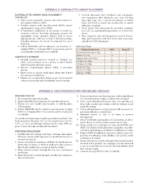
APPENDIX D: SUPRAGLOTTIC AIRWAY PLACEMENT

              SUPRAGLOTTIC AIRWAY (SGA) PLACEMENT                  •  Confirm placement with ventilation and auscultation
              CHECKLIST                                              over epigastrium, then bilaterally over chest, left lung
                •  Open airway manually, measure and insert simple air-  then  right  lung.  Get  a  second  practitioner  to  double
                  way adjunct (NPA or OPA).                          check and verify in sounds are questionable or cannot
                •  Ventilate patient with bag-valve-mask (BVM) (attach   otherwise auscultate.
                  supplemental oxygen, if available).              •  Verify proper SGA placement by secondary confirma-
                •  If ventilations insufficient, or the patient is clearly un-  tion such as capnography/capnometry or colorimetric
                  conscious and not breathing adequately, prepare for   device.
                  supraglottic airway insertion. Inspect SGA to ensure   •  Place orogastric tube and decompress stomach if avail-
                  appropriate size. Lubricate airway to facilitate passage.   able, and compatible with SGA device (has a port spe-
                  Cricothyroidotomy  kit  should  be  prepared  for  use  if   cifically for OGT placement).
                  SGA fails.
                •  Follow MSMAID and for induction, use ketamine (1–  SGA Size Chart
                  2mg/kg IV/IO or 3–4mg/kg IM) if time permits and the   Estimated patient size  LMA   King LT*
                  recommended medications are available.         Neonates/infants (up to 5kg)  1          0
                                                                 Infants 5–10 kg              1.5         1
              INSERTING THE AIRWAY
                •  Properly position head in a neutral or “sniffing” po-  Infants/children 10–20kg  2     2
                  sition (neck extended, as on a pillow or small blanket   Children 20–30kg   2.5         2.5
                  while lying flat) and open airway.             Children 30–50kg             3           3*
                •  Remove oropharyngeal airway (OPA) if previously   Adults 50–70kg           4           4*
                  placed.                                        Adults 70–100kg              5           5*
                •  Insert device to proper depth (may adjust later if need   Adults > 100kg   6
                  for improved ventilation).
                •  Inflate cuff, if applicable; inflate as per device-specific
                  volume instructions and immediately remove syringe.


                                  APPENDIX E: CRICOTHYROIDOTOMY PROCEDURE CHECKLIST

              PREPARE PATIENT                                    5.  Open and maintain membrane incision with tracheal hook
              1.  Pre-oxygenate patient if possible.                (or curved hemostat, bougie, or blunt end of scalpel).
              2.  Inspect/assemble/test equipment for cricothyroidotomy.  6.  Insert endotracheal/tracheostomy tube into opening and
              3.  Prepare site with alcohol and betadine or chlorhexadine   direct tube caudad into trachea until the balloon is just
                (Chlora-prep).                                      inside the airway.
              4.  Follow MSMAID and for induction, use ketamine (1mg/kg   7.  Inflate cuff and detach syringe (palpate bulb to ensure it is
                IV/IO or 3–4mg/kg IM) if time permits and the medication   not underinflated or overinflated).
                is available.                                    8.  Maintain  control  of  tube  at  all  times  to  prevent
                                                                    dislodgement.
              For awake cricothyrotomy: Explain procedure to patient; *Use
              local anesthesia: lidocaine (1% or 2%), bupivacaine (0.25%,   9.  Attach waveform capnography, or capnometry, or colori-
              0.5%, or 1%); local through planned incision area AND ap-  metric device to confirm proper placement of tube.
              prox. 1–2mL through cricothyroid membrane          10.  Being careful not to dislodge the tube, attach BVM with
                                                                    PEEP and further check placement (epigastric and bilat-
              PERFORM PROCEDURE                                     eral chest) and adequacy of bilateral insufflation of lungs.
              1.  Stabilize thyroid cartilage and keep overlying skin taught.   11.  Remove BVM (if sufficient respiratory effort), assess res-
                Maintain control with hand until the membrane incision is   pirations for adequacy (rate, rhythm, and quality), assist
                secured (see step 8).                               ventilations if needed.
              2.  Locate cricothyroid membrane (Palpate for hyoid and tra-  12.  Secure with sutures and tie with girth hitch passed around
                cheal rings. If unsure or difficult landmarks, then measure   the neck if time permits. As a stopgap, may use chest seal
                three finger widths above sternal notch for adults.)  or secure around the neck with tie, ensuring inflation bulb
              3.  Make vertical incision through the skin over cricothyroid   does not get caught.
                membrane.                                        13.  Consider placing NG/OG tube if available.
              4.  Make horizontal incision through cricothyroid membrane,
                then immediately:


                                                                                        Airway Management in PFC  |  151