
Page 154 - JSOM Fall 2020
P. 154
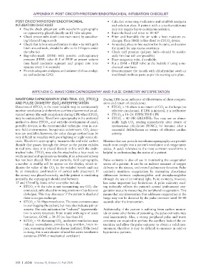
APPENDIX F: POST CRICOTHYROTOMY/ENDOTRACHEAL INTUBATION CHECKLIST

          POST CRICOTHYROTOMY/ENDOTRACHEAL                     •  Calculate remaining medication and establish analgesia
          INTUBATION CHECKLIST                                    and sedation plan. A patient with a cricothyroidotomy
            •  Double check placement with waveform capnography   may not require heavy continuous sedation.
               or capnometry, placed directly on ET tube adapter.  •  Raise the head and torso to 30–45°
            •  Check proper tube depth (not main stem) by auscultat-  •  Filter and humidify the air with a heat moisture ex-
               ing bilateral lung sounds                          changer. Place HME in-line distal to ETCO  device.
                                                                                                   2
            •  Check that tube is secured (suture to skin + tie with girth   •  As needed, place in-line suction for the tube, and suction
               hitch around neck, should be able to fit 2 fingers under   the mouth for any excess secretions
               the tube tie)                                   •  Check cuff pressure (palpate bulb—should be moder-
            •  Bag-valve-mask (BVM) with positive end-expiratory   ately firm but still compressible)
               pressure (PEEP) valve @ 5 of PEEP at proper volume   •  Place orogastric tube, if available.
               (one hand moderate squeeze) and proper rate (one   •  Put a BVM + PEEP valve at the bedside if using a me-
               squeeze every 5–6 seconds)                         chanical ventilator.
            •  Provide adequate analgesia and sedation (follow analge-  •  Decontaminate the mouth with chlorhexidine swab or
               sia and sedation CPG)                              toothbrush without paste as per the nursing care plan.


                    APPENDIX G: WAVEFORM CAPNOGRAPHY AND PULSE OXIMETRY INTERPRETATION

          WAVEFORM CAPNOGRAPHY (END-TIDAL CO  [ETCO ])       During CPR (as an indicator of effectiveness of chest compres-
                                                  2
                                                        2
          AND PULSE OXIMETRY (SpO ) INTERPRETATION           sions and return of circulation):
                                   2
          Detection of ETCO  is the most reliable way to continuously   •  ETCO  < 10: there is no return of CO  to the lungs (no
                                                                                                2
                                                                       2
                         2
          monitor ventilation and therefore confirm placement of an ad-  effective circulation). If CPR is initiated, it is ineffective
          vanced airway (the only exception is during CPR when ETCO    •  ETCO  = 10–20: EFFECTIVE CPR
                                                                       2
                                                         2
          may be undetectable). Waveform capnography is the preferred   •  ETCO  = 40 OR GREATER: You may see an abnor-
                                                                       2
          method to detect ETCO , and with the development of small,   mally high CO  reading immediately after return of
                             2
                                                                              2
          portable devices, is the recommended technique even in aus-  spontaneous circulation (ROSC), for instance after a
          tere field environments. Inexpensive colorimetric CO  detec-  successful defibrillation or return of effective cardiac
                                                    2
          tors are available; however, the color change method may be   activity.
          very difficult to visualize with poor lighting or night vision de-
          vices. Waveform capnography measures the end-tidal carbon   Monitor that can provide waveform capnography can provide
          dioxide that passes through the device as the patient exhales   much more insight into a patient’s ventilation and oxygenation
          in real time since it is placed directly in-line with the endo-  status. A quick reference to the most common waveforms is
          tracheal tube. ETCO  may also be attached to a face mask to   helpful to understanding the status of a patient.
                          2
          verify normal and spontaneous breaths, if an advanced airway
          has not been placed. With most portable, field capnographs,   Pulse oximetry is also of use in monitoring the oxygenation
          a number in mmHg will be appear on the display, which in-  status of a patient. It can be an indirect measure of oxygen
          dicates the value of the CO  in the exhaled breath and can   delivery to the tissues, and overall pulmonary function. Pulse
                                2
          be an immediate confirmation of correct tube placement. If   oximetry monitors oxygenation by measuring absorbance
          the airway was placed correctly, and the patient is ventilating   differences  between  oxyhemoglobin  and  deoxyhemoglobin
          normally, the capnograph should read between       through the use of an infrared light. Pulse oximetry, however,
          35 and 45mmHg. Some other examples include:        has some important key limitations. A pulse oximetry read-
            •  ETCO  = 0: the tube is not transmitting any CO : dis-  ing indirectly reflects the patient’s central (pulmonary) oxy-
                                                     2
                    2
               connected, tube placed in wrong position or has become   genation status by measuring the peripheral oxygenation. This
               dislodged. This may also occur if the patient is dead and   means that any intervention that addresses oxygenation in the
               there is no gas exchange.                     lungs may not be detected by the pulse oximeter until 30–90
            •  ETCO  < 35: Hyperventilation. The most common cause   seconds after the intervention.
                    2
               is over-bagging the patient, but may also indicate pain or
               anxiety. The only indication for “induced” hyperventila-  Additionally, if the patient is suffering from carbon monox-
               tion is severe traumatic brain injury with signs of acute   ide or some other forms of poisoning, the pulse oximetry may
               herniation, GOAL = 30–35 (no less than 30)    read inaccurately. Also, a strong peripheral pulse and warm
            •  ETCO  > 45: Retaining CO , ineffective ventilation may   extremity are required to perfuse the capillary beds of the ex-
                    2
                                    2
               indicate oversedation, primary lung problem, brain in-  tremities and allow the pulse oximeter to obtain a valid mea-
               jury, worsening obstructive disease (asthma). If the trend   surement, therefore it may be difficult to measure in cold or
               is rising, this is an indicator of need for active ventilation   hypotensive patients.
               assistance (BVM or mechanical ventilator)


          152  |  JSOM   Volume 20, Edition 3 / Fall 2020