
Page 72 - JSOM Spring 2020
P. 72
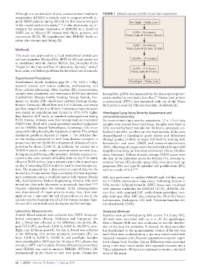
Although not yet standard of care, extracorporeal membrane   FIGURE 1  Altitude exposure profile of each day’s experiment.
          oxygenation (ECMO) is already used to support severely in-
          jured ARDS patients during AE and for that reason was part         Pre flight – Vitals, Labs
          of the model used in this study. 14–16  In this pilot study, we in-
          vestigate the systemic expression of HMGB1 in a model of
          ARDS due to bilateral PC treated with fluids, pressors, and         5,000 – 30 minutes
          venovenous ECLS. We hypothesized that HMGB1 levels in-           Vitals, Labs @ 15 min, 30 min
          crease after trauma and during AE.
                                                                               8,000 – 30 minutes
          Methods                                                          Vitals, Labs @ 15 min, 30 min
          This study was approved by a local institutional animal care
          and use committee (Protocol No. BPTS 15-02) and carried out         30,000 – 15 minutes
          in compliance with the Animal Welfare Act, principles of the
          “Guide for the Care and Use of Laboratory Animals,” and all
                                                                              5,000 – 15 minutes
          local, state, and federal guidelines for the ethical use of animals.  Vitals, Labs @ 15 min
          Experimental Procedures
          Anesthetized, female Yorkshire pigs (N = 15, 53.8 ± 1.4kg)         Post flight – Vitals, Labs
          received arterial and venous catheters, tracheostomy, and
          Foley catheter placement. After baseline (BL) measurements,
          animals were cannulated and venovenous ECLS was initiated   hemoglobin (pfHb) was measured via the direct spectrophoto-
          (CardioHelp; Maquet Gmbh, Gettinge Group, Rastatt, Ger-  metric method as previously described.  Plasma total protein
                                                                                           19
          many) via Avalon 23Fr dual-lumen catheter (Getinge Group,   concentration  (PTPC)  was  measured  with  use  of  the  Pierce
          Rastatt, Germany). Blood flow was 1.2–2.2L/min, and sweep   BCA protein assay kit (Thermo Scientific, Rockford, IL).
          gas flow ranged from 4 to 8L/min. Continuous heparinization
          was started at cannulation and titrated to 30%–50% higher   Histological Lung Injury Severity Assessment and
          than baseline ACT levels, as standard anticoagulation during   Immunohistochemistry
          ECLS therapy. Animals were then transported via a standard   For postmortem injury severity assessment, 1.5 × 1.5-cm lung
          NATO litter fitted with a next-generation medical equipment   samples were excised from both lungs. Samples were fixed in
          rail kit (MERK; Smeed Technologies, Cummings, GA) to an   10% normal buffered formalin for 48 hours, processed, em-
          adjacent building housing the hypobaric chamber. The altitude   bedded in paraffin, and then cut into 4µm sections. Slides were
          simulation profile is depicted in Figure 1. The altitudes cho-  deparaffinized in histological grade xylene and dehydrated
          sen for testing correspond to both long-distance transport in   through graded alcohols to water, followed by staining with
          pressurized aircraft (8,000 ft) and potential altitudes of trans-  hematoxylin and eosin (H&E) and immunohistochemistry
          portation by drone (5,000 ft). In addition, we carried out a   (IHC). Histological images were obtained with ×100 and ×200
          30,000-ft step to study a rapid decompression scenario (e.g.,   magnification using an Axioskop microscope (Zeiss, Oberko-
          during an aircraft losing cabin pressure). Altitude exposure oc-  chen, Germany). Diffuse alveolar damage (DAD) scores were
          curred in the same animals in healthy state on day 1 (to study   the sum of the individual scores for fibrosis (%), alveolar in-
          effects of ECLS without injury present) and in the injured state   terstitial fibrosis (IF), alveolar space (AS), alveolar protein ag-
          on day 2 (assuming ECLS would be used to treat trauma vic-  gregation (PA) and type II epithelial cell proliferation (EC),
          tims. The uninjured day 1 data served as control data for the   each on a scale of 0–4. 20–22
          injured day 2 experiments. Injury consisted of bilateral pulmo-
          nary contusions using a modified captive-bolt stunner (Model   IHC was performed to analyze HMGB1 and Toll-like recep-
          ML; Karl Schermer, Packers Engineering, Omaha, NE) with   tor 4 (TLR4) expression in lung tissue. Following fixation in
          immediate chest-tube placement as previously described. 7,17,18    10% normal buffered formalin (NBF), tissue was incubated
          Heparin administration for systemic ECLS anticoagulation   with primary antibodies for HMGB1 (1:150, ab18256; Ab-
          was discontinued ~8 hours prior to injury; thus, from mid-  cam Inc.) with standard IHC avidin-biotin-peroxidase com-
          night of day 1 and until the end of procedures on day 2, the   plex technique (Elite ABC kits, Catalog No. PK-6100; Vector
          animals received heparin-free ECLS for trauma because hepa-  Laboratories, Burlingame, CA) with 3-diaminobenzidine tet-
          rin would be contraindicated during trauma/hemorrhage.  rahydrochloride (DAB).

          Laboratory Measurements                            Statistical Methods
          Arterial blood samples were collected into EDTA blood-col-  Statistics were performed using SAS version 9.4 (Cary, NC).
          lection vacutainers (Becton, Dickinson and Company). On   All tests were two-sided with an  α = .05 for significance.
          day 1, blood was collected at the following time points: BL,   First a Shapiro-Wilk test was conducted to test the distribu-
          post-ECLS (PE), sea level, 5,000 ft, 8,000 ft, 30,000 ft, post-  tion of the data for normality. If skewed, the data were then
          flight, and 12 hours post-PE. On day 2, blood was collected   log transformed or the nonparametric version of the test was
          at the following time points: preinjury, postinjury (PI), sea   used. Data were analyzed using a one-way mixed model with
          level, 5,000 ft, 8,000 ft, 30,000 ft, and postflight. Samples   repeated measures and a Dunnett adjustment to test for signif-
          were centrifuged at 3000 rpm for 10 min at 4°C; plasma was   icant change from baseline. Group differences were examined
          stored at –80°C until analysis. Enzyme-linked immunosorbent   using a two-way mixed model with repeated measures and a
          assay (ELISA) was used to measure HMGB1 (ST51011; IBL   Tukey adjustment. All data are expressed as means ± standard
          International) in the blood at each time point. Plasma-free   error of the mean.


          66  |  JSOM   Volume 20, Edition 1 / Spring 2020