
Page 73 - JSOM Spring 2020
P. 73
Results FIGURE 3 DAD component scoring in right and left lungs.
Representative images of the right (A) and left (B) lungs (hematoxylin
Of 15 animals that entered the study, all completed day 1 (con- and eosin stain; original magnification ×200). (C) Injury scores for
trol conditions). Six animals died after PC but before flight on alveolar interstitial fibrosis (IF), alveolar air space (AS), protein
day 2: two died from suspected myocardial infarction follow- aggregate (PA), and expression of type II alveolar epithelial cell (EC).
ing injury and four died from post-PC cardiac contusion and (D) Percentage of tissue fibrosis and DAD score. Values are means ±
standard error.
nonresponsiveness to vasopressors and fluids, signifying the
severity of the model.
On gross observation, severe lung laceration, damage to major
blood vessels, and severe contusion resulted in bilateral lung
damage (Figure 2). Histological scoring of lung injury (diffuse
alveolar damage [DAD]) is shown in Figure 3A (left lung) and
3B (right lung) for both left and right sides. Interstitial fibrosis
(IF), alveolar space (AS), protein aggregation (PA), and type
II epithelial cell (EC) proliferation scores were not different
between right and left lungs (Figure 3C). The fibrosis percent-
age (15.7% ± 1.8% versus 15.0% ± 1.9%) and overall DAD
scores (22.8 ± 2.4 versus 21.5 ± 2.2) were not different be-
tween lungs, an indication of identical injury to both lungs
(Figure 3D).
FIGURE 2 Postmortem gross anatomy images of posterior lungs
(A) and anterior lungs (B) after bilateral pulmonary contusion
supported by ECLS. Arrows point to areas of consolidation as a
result of trauma.
(A) (B)
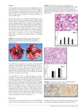
Extracellular and paranuclear cytoplasmic immunochemical
staining revealed HMGB1 and TLR4 in the lungs of all ani-
mals (Figure 4A, B). These mediators were primarily localized
in the alveolar epithelial cells and neutrophils, monocytes,
macrophages, and endothelium; however, there were no dif-
ferences in the expression pattern/area or density of HMGB1
and TLR 4 in right versus left lungs.
Plasma HMGB1 on day 1 was transiently higher at arrival to
the altitude chamber (D1 sea level time point) (Figure 5A).
HMGB1 increased significantly after injury reaching highest
values at 8,000 ft on day 2, after which levels decreased but re-
mained elevated when compared to baseline (Figure 5A). pfHb FIGURE 4 Postmortem expression of (arrows) HMGB1 (A) and
decreased significantly on day 1 after ECLS initiation (PE time TLR4 (B) after bilateral pulmonary contusion.
point), at 30,000 ft. and at the 12-hour time point (Figure
5B). On day 2, pfHb was higher at all time points but statis-
tically significantly higher at 8,000 ft and postflight. Plasma
total protein concentration (PTPC), a nonspecific measure of
protein breakdown, was significantly decreased from day 1
sea level until the end of the experiment (Figure 5C), likely
reflecting hemodilution due to resuscitation.
Discussion
This is the first report on the systemic expression of HMGB1 with ECLS. We found that HMGB1 increased with trauma
using a model of combat-relevant injury and AE managed and remained elevated at subsequent stages of the study.
HMGB1 Protein Expression in a Polytrauma Model | 67