
Page 17 - JSOM Summer 2019
P. 17
The rash blanched with pressure. Additional symptoms in- skin eruptions may precede retiform purpura in medical condi-
cluded shortness of breath and generalized malaise. A physical tions to include calciphylaxis, antiphospholipid antibody syn-
examination revealed left lower leg paresis. drome, and warfarin-induced skin necrosis. According to the
3
Divers Alert Network Annual Diving Report 2017 Edition,
He was immediately placed on surface oxygen on the diving there were 76 cases of cutis marmorata in 2015. However,
boat and then transported to the state hospital via ambulance. the true incidence is unknown because there are no official
He was treated with surface oxygen at 11 L/min overnight via reporting requirements and because cases may not be reported
nonrebreather mask. His symptoms improved on surface oxy- to the Divers Alert Network Medical Services Call Center
gen, which he continued to breathe until transport was secured. (DANMSCC), which provides medical information in addi-
tion to coordinating treatment and evacuation for injured div-
He was appropriately transported to Guam via turboprop air ers. Well known to the US Navy and civilian divers, the Divers
ambulance with the cabin pressurized to 1 standard atm of Alert Network collects data regarding diving injuries, not in-
pressure and at 2133.6 m of altitude. He arrived at the Na- cluding fatalities, from DANMSCC and the Annual Survey of
val Base Guam Recompression Chamber about 36 hours af- Hyperbaric Chambers. The initial information is entered into
ter the onset of symptoms. By this time, shortness of breath a database. Follow-up is conducted to confirm actual diagno-
had resolved, and the left lower leg demonstrated movement sis of DCS. Results are analyzed and published in the Annual
against some resistance on examination. The skin changes had Diving Report. 4
remained confined to the chest and abdomen but had become
erythematous. DCS is a pathologic response to bubble formation. The patho-
physiology is governed by Henry’s law: the amount of gas that
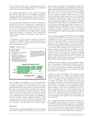
The treating team decided to pursue hyperbaric recompression will dissolve in a liquid at a given temperature is directly pro-
according to a Treatment Table 6 protocol with maximum ex- portional to the partial pressure of that gas. Inert gas, mainly
tensions at 18 m. Figure 3 from the US Navy Diving Manual nitrogen, is forced into the tissues of the body by increased
depicts the treatment protocol. 2 ambient pressure at depth during dives. During controlled as-
cent, inert gas gradually comes out of solution as ambient pres-
FIGURE 3 Treatment Table 6. 2 sure decreases. When ambient pressure reduction occurs too
quickly, intravascular and extravascular bubbles may form.
1
The latter is easily visualized when opening a beer can. Remov-
ing the lid causes a quick reduction in pressure and bubbles
form as a result. In this case, the diver omitted more than 300
minutes of decompression time, which is time expected to be
spent at depth allowing nitrogen gas to slowly come out of his
tissues. This severely increased his risk of DCS.
Bubbles cause injury through two main mechanisms: mechan-
ical and nonmechanical. Mechanically, bubbles cause distor-
tion or compression of tissue, leading to pain and edema, or
it can cause vascular obstruction, leading to stroke-like signs.
Nonmechanically, bubbles may act as a foreign body, provok-
ing the inflammatory response. 1
A literature search reveals multiple cutis marmorata images
of deeply erythematous and violaceous skin eruptions that are
similar to the skin changes of this patient at 36 hours after
onset. The mechanisms of injury support the transient nature
of cutaneous DCS lesions. Vascular obstruction initially leads
He was brought to 18 m of depth at a rate of 6.096 m/min. He to ischemia and, therefore, early reticular, violaceous changes.
breathed 100% oxygen for three 20-minute periods at 18 m Eventually, the inflammatory response causes deep erythema.
according to standard protocol. His treatment was extended However, there are limited data characterizing DCS skin le-
with two additional 20-minute periods at 18 m as his left lower sions in relation to time. One study published in July 2017
leg weakness resolved toward the end of the third oxygen pe- depicts a temporal development of skin lesions in swine with
riod. All oxygen periods at 18 m were followed by 5-minute induced DCS. The lesions were characterized from stage I to
air breaks to prevent oxygen toxicity. He continued to breathe stage VI. The reticular lesions depicted in Figures 1 and 2 in
100% oxygen during a 30-minute ascent at 0.3048 m/min to this case are comparable to lesions of stage I and stage II in the
9 m, at which time a 15-minute air break was administered. study, respectively, which appeared early after surfacing and
The table concludes with two additional 60-minute, 100% were short lived. 5
oxygen periods at 9 m with an interval 15-minute air break
between periods. The patient was then brought back to sur- DCS is categorized into type I and type II. Type I DCS is fur-
face, ascending at a rate of 0.3048 m/min, on 100% oxygen. ther classified as musculoskeletal, cutaneous, or lymphatic.
2
Type II DCS is further classified as pulmonary or neurological.
Neurological DCS is further subclassified into peripheral, spi-
Discussion
nal cord or central nervous system, cerebral, vestibular, and
Cutis marmorata in the adult population is rarely associated ocular. Characteristics of the subtypes of type I DCS and type
with medical conditions other than DCS. Violaceous, reticular II DCS are delineated in Table 1 and Table 2, respectively. 1
Skin Changes After Aggressive Diving | 15