
Page 132 - JSOM Summer 2019
P. 132
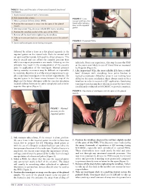
TABLE 2  Steps and Principles of Improvised Inguinal Junctional
          Tourniquets
           1. Apply manual pressure with a hemostatic.
           2. Sole rescuers take a knee.
                                                             FIGURE 2  Lone
           3. Select a pressure delivery device (PDD).       rescuer with two free
           4.  Position the tourniquet or strap over the apex of the gluteal   hands after taking a
            muscle.                                          knee in the inguinal
                                                             gutter.
           5. Like improvised TQs, the most reliable IJTs have a windlass.
           6. Position the windlass medial of the apex of the PDD.
           7. Remove all the slack before tightening the windlass.
           8.  Take up tourniquet slack in a pushing motion across the patient’s
            body.
                                                             FIGURE 3  Potential PDDs.
           9. Junctional tourniquets move during transport.


            followed by either a knee or a fist placed squarely in the
            inguinal gutter on the injured side. Pack the wound with
            an approved hemostatic followed by direct pressure. This
            step is crucial and can afford the casualty precious time
            while tourniquet preparations are made. Faltering on this   inferiorly. From our experience, this step focuses the PDD
            principle may result in the exsanguination of the casualty   at the point most likely to cut off blood flow as measured
            prior to application of the tourniquet. Manual pressure   by Doppler (Figure 4).
            during casualty movement may be difficult or impossible   5.  Like improvised TQs, the most reliable IJTs have a wind-
            to maintain, therefore it is of the utmost importance to ap-  lass.   Attempts with everything from pelvic binders to
                                                                   5
            ply a junctional tourniquet at the earliest opportunity. The   tactical compression (Murphy) wrap in our training have
            inguinal gutter is the linear crease between the top of the   all led to the same conclusion: there always needs to be a
            thigh and the lower abdomen with the vascular structures   windlass in order to succeed at IJT application. Ratcheting
            located midway between the pubic symphysis and anterior   tourniquet mechanisms have shown promise but have been
            superior iliac spine (Figure 1).                   insufficiently evaluated at SOCMSSC to provide comment.
                                                             FIGURE 4  Placement of tourniquet over the apex of the gluteal
                                                             muscle.


                                           FIGURE 1  Manual
                                           pressure in the
                                           inguinal gutter.


          2.  Sole rescuers take a knee. If the rescuer is alone, prefera-
            bly use a knee in the inguinal gutter in order to have two   6.  Position the windlass (depicted by red line) slightly medial
            hands free to prepare the IJT. Palpating distal pulses or   of the apex of the PDD. This discovery was made during
            with the use of a Doppler as described later can refine the   the many thousands of repetitions at IJT training during
            best pressure location of this technique. If pelvic fracture is   SOCMSSC, especially with cylindrical or curved PDDs.
            suspected, the rescuer must weigh the importance of hem-  These observations were made purely by trial and error
            orrhage control over the potential for further harm through   and not through any structured research processes. This
            manipulation of a fractured pelvis (Figure 2).     step most consistently directs the PDD toward the femoral
          3.  Select a PDD. An object that fits into the inguinal gutter   artery and prevents it moving more posteriorly compared
            and can remain stable is half of the solution. The object   to pressure directly over or lateral to the apex (Figure 5).
            will usually be something either cylindrical or spherical.   7.  Remove all the slack before tightening the windlass. Failure
            Munitions or pyrotechnic devices should never be used as   to take up all the slack is the number one reason for tour-
            the PDD (Figure 3).                                niquet failure.
          4.  Position the tourniquet or strap over the apex of the gluteal   8.  Take up tourniquet slack in a pushing motion across the
            muscle. The apex of the gluteal muscle is the top part of   patient’s body. Tourniquet slack can be difficult to take up
            the buttocks and this prevents the strap from sliding off   when pushing a small amount of strap into the ground or a


          130  |  JSOM   Volume 19, Edition 2 / Summer 2019