
Page 60 - JSOM Winter 2018
P. 60
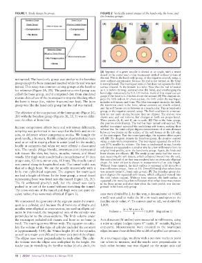
FIGURE 1  Study design by group.                   FIGURE 2  Vertically paired images of the band-only, the bone, and
                                                             the boneless groups.
                                                              (A)             (C)             (E)


                                                              (B)             (D)             (F)


                                                             (A)  Segment of  a green  noodle  is  shown at  an angle,  with a  wood
                                                             dowel at the center and a blue tourniquet applied without a turn of
                                                             the rod. This is the band-only group, or the negative control, setup, a
          not turned. The band-only group was similar to the boneless   state without compression because the rod is not turned, so the band
          group except the bone remained inserted while the rod was not   is not tightened. The tourniquet rests on but does not squeeze the limb
          turned. This setup was common among groups as the baseline   surface inward. At the bottom is a ruler. Note that the rod is located
          for reference (Figure 2A, 2B). The positive control group was   as it is before turning: centered over the band, and overhanging the
          called the bone group, and it comprised data from simulated   band edge transversely by 2–2.125 inches. Users of this visual cue can
          routine clinical use of the tourniquet to stop the bleeding when   gauge if the band is 2–3 inches above the wound. (B) The diagram de-
                                                             picts the limb end-on. In cross-section, the limb under the tourniquet
          the bone is intact (i.e., neither fractured nor lost). The bone   includes soft tissues and bone. The blue tourniquet encircles the limb,
          group was like the band-only group but the rod was turned.  the innermost circle is the bone, whose contents are manila colored,
                                                             and the soft tissues are in between in a peach color. This is band-only
                                                             group, or the negative control, setup. The limb and bone in cross-sec-
          The objective of the comparison of the bone group (Figure 2C,   tion are not quite circles, because they are ellipses. The end-on view
          2D) with the boneless group (Figures 2E, 2F, 3) was to delin-  shows area and not volume, but changes in both are proportional.
          eate the effect of bone loss.                      Three panels (B, D, and F) are to scale. (C) This is the bone group,
                                                             the positive control setup. The rod has been turned and secured. The
          Because compression affects bone and soft tissues differently,   applied tourniquet squeezed the underlying soft tissues, making their
          sampling was performed in two ways for the limb and its tis-  volume less. An outer caliper depicts measurement of an axis distance
                                                             between two points on the surface of the soft tissues at the left edge
          sues, to delineate where compression works. We bought the   of the tourniquet. Past the tourniquet edge, the squeeze effect tapers
          pool noodle, a buoyant, flexible cylinder of polyethylene foam   off. (D) The diagram depicts the boneless group, the positive control
          used as an aid in staying afloat in water (used for the model),   setup, a state with compression. The tourniquet squeezed the soft tis-
          locally at summer’s end when we were offered a discounted   sues 17% smaller by volume. The bone is unchanged in size, but the
                                                             soft tissues are squeezed to a smaller size by a few millimeters from its
          rate. The noodle (Mega Noodle, swimways.com) represented   original limb profile, as in the band-only group. Again, the limb and
          soft tissues like skin, fat, fascia, muscles, nerves, and blood   bone shapes are ellipses. (E) The study group setup had the tourni-
          vessels. The thigh-wide noodle had a circumference of 37.1cm   quet squeezing the soft tissues, which collapsed inward into the void,
          (major axis, 121mm; minor axis, 113mm). The noodle tunnel   the central tunnel of air that was rendered into an obviously elliptical
          was central along its longitudinal axis. The tunnel width was   shape. An inner caliper is shown in measurement of an axis length.
                                                             Without bone support, the limb surface is squeezed a bit more by a
          that of a thigh bone. We cut the noodle transversely with a   few millimeters (mean, 3mm or 3% [3mm/103mm]) than when bone
          knife  into cylindrical  segments.  The  segment  for tourniquet   was present (panel C, band-only group). (F) The boneless group dia-
          use had a height of 65mm. In the bone group, a wood dowel   gram depicts the squeezed soft tissues, which collapsed inward into
          representing bone was fitted into the tunnel (Figure 2A, 2C).   the void (white region). Without bone support, the limb surface is
                                                             squeezed a bit more by a few millimeters than when bone was present
          The fit withstood gravity’s pull, but the dowel was easily   in the bone group and also than when the limb profile was uncom-
          pushed in or out of the tunnel without stretching the tunnel.   pressed in the band-only group.
          The cross-sections of the tunnel and thigh were not quite cir-
          cular; rather, they were truly elliptical (Figure 3).  axes were divided by 2. In this way, a denominator of 4 (M/2
                                                             × m/2) was used to make the M × m result analogous to the
                                                                                           3
          We represented the geometry of the segment under the tourni-  familiar circle value, r . To convert mm  to mL, we divided by
                                                                              2
          quet as a cylinder, and because the dimensions of thighs and   1,000:
          noodles were elliptical in cross-section, we used an elliptic cyl-  (π × M × m × h)
          inder. In this model, the longitudinal axis of the cylinder was      Volume =    4   / 1,000   (Equation 1)
          perpendicular to the cross-sections. The limb volume under
          the tourniquet included soft tissues and bone or no bone (a   Axis distances (M and m) were measured in millimeters, using
          void). The tourniquet was 40mm wide. The equation to calcu-  a ruler or caliper (caliper types: 8" inside, 8" outside; bighorn
          late the volume of this type of cylinder included the constant   corp.com). Measurements were oriented to the tourniquet
          π (approximately 3.14), the 40mm height (h) of the cylinder,   edges because those defined the width of applied compression.
          as well as a major axis (M) and a minor axis (m) of the ellipse.
          The ellipse axes were perpendicular to each other. The cylin-  At a tourniquet edge, we drew with a pen two marks to point
          der volume was the ellipse area multiplied by the height. For   out where to measure, and the marks were perpendicular to
          reader ease in mimicking the familiar radius (r) of a circle, the   each other because one was aligned on the major axis and


          58  |  JSOM   Volume 18, Edition 4 / Winter 2018