
Page 126 - JSOM Winter 2018
P. 126
preformed yet unstable thrombus. Particular to the OpK9, re- up to 85% of human trauma patients remain in a state of mi-
main cognizant of external wounds and bleeding that remain crocirculatory shock even after normalization of traditional
hidden by the canine’s hair coat. macrohemodynamic parameters. 16,17
A Focused Assessment with Sonography in Trauma (FAST)
Identifying Hemorrhage
examination is a valuable tool for quickly and reliably iden-
Internal Hemorrhage tifying intracavitary hemorrhage in canines, as in humans. 18–20
Identifying life-threatening, trauma-induced, internal hemor- Point-of-care ultrasound is becoming increasingly more acces-
rhage in canines is essentially the same as in humans. With- sible at the point of injury on the battlefield and in the civilian
out the use of advanced imaging modalities, early diagnosis prehospital environment. 18–20 The technique for performing a
of intracavitary hemorrhage is often challenging, especially FAST examination in canines is similar to that of humans; a
when there are concurrent trauma- and shock-induced factors brief description follows. 18,21
such as altered mental status and extra-abdominal injuries
(e.g., neurologic injuries). In the field, recognition of intracav- 1. Place the canine in right (preferable due to cardiac notch)
itary hemorrhage is primarily based on the history of blunt or or left lateral recumbency. In the presence of respiratory
penetrating trauma along with physical examination findings distress, scan the canine while it is standing.
and alterations in vital signs indicative of hemorrhagic shock Important: Avoid placing a critically injured canine in
(e.g., mental depression, tachycardia, prolonged capillary refill dorsal recumbency, because of the increased risk for com-
time, pale mucous membranes, poor pulse quality, and cold promising respiratory function and hemodynamics. Addi-
extremities) 10–12 (Table 1). tionally, FAST fluid scoring systems were only validated
with canines in lateral recumbency. 21
TABLE 1 Operational Canine Vital Signs: Perfusion Parameters 2. Shaving the hair coat is not required. To ensure adequate
HR , probe-to-skin contact, apply isopropyl alcohol and/or
a
per Pulse SBP, acoustic coupling gel to the hair coat before probe place-
Stage of Shock min CRT, s MM Mentation Quality mmHg ment. Note: Contact the manufacturer to ensure isopropyl
Normal Bright, alcohol will not damage the probe head.
(at rest) <120 <2 Pink alert Strong >90 3. Abdominal FAST (AFAST): involves scanning the abdomen
Acute >120 <1 Red Normal Fair >90 at the following four sites (Figure 1):
compensatory
Early >140 >2 Pale Depressed Weak <90
decompensatory
Terminal <80 Absent Pale Obtunded Absent Low
CRT, capillary refill time; HR, heart rate; MM, mucous membrane; SBP,
systolic blood pressure.
a Exercise or other activity, pain, and stress will result in higher HR.
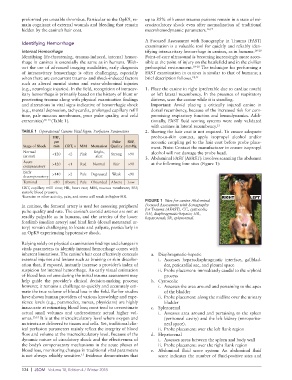
FIGURE 1 Sites for canine Abdominal
In canines, the femoral artery is used for assessing peripheral Focused Assessment with Sonography
pulse quality and rate. The canine’s carotid arteries are not as for Trauma (AFAST). CC, cystocolic;
DH, diaphragmatic-hepatic; HR,
readily palpable as in humans, and the arteries of the lower hepatorenal; SR, splenorenal.
forelimb (median artery) and hind limb (dorsal metatarsal ar-
tery) remain challenging to locate and palpate, particularly in
an OpK9 experiencing hypotensive shock.
Relying solely on physical examination findings and changes in
vitals parameters to identify internal hemorrhage comes with
inherent limitations. The canine’s hair coat effectively conceals a. Diaphragmatic-hepatic
external injuries and lesions such as bruising or skin discolor- i. Accesses hepatodiaphragmatic interface, gallblad-
ation that, if exposed, instantly increase a provider’s index of der, pericardial sac, and pleural space
suspicion for internal hemorrhage. An early visual estimation ii. Probe placement: immediately caudal to the xiphoid
of blood loss volume during the initial trauma assessment may process
help guide the provider’s clinical decision-making process; b. Cystocolic
however, it remains a challenge to quickly and accurately esti- i. Assesses the area around and pertaining to the apex
mate the true volume of blood loss in the field. Earlier studies of the bladder
have shown human providers of various knowledge and expe- ii. Probe placement: along the midline over the urinary
rience levels (e.g., paramedics, nurses, physicians) are highly bladder
inaccurate at estimating blood loss; most tend to overestimate c. Splenorenal
actual small volumes and underestimate actual higher vol- i. Assesses area around and pertaining to the spleen
umes. 13,14 It is at the microcirculatory level where oxygen and (peritoneal cavity) and the left kidney (retroperito-
nutrients are delivered to tissues and cells. Yet, traditional clin- neal space).
ical perfusion parameters mainly reflect the integrity of blood ii. Probe placement: over the left flank region
flow and volume at the macrocirculatory level. Because of the d. Hepatorenal
dynamic nature of circulatory shock and the effectiveness of i. Assesses areas between the spleen and body wall
the body’s compensatory mechanisms in the acute phases of ii. Probe placement: over the right flank region
blood loss, monitoring changes in traditional vital parameters e. Abdominal fluid score system: An abdominal fluid
is not always reliably sensitive. Evidence demonstrates that score indicates the number of fluid-positive sites and
15
124 | JSOM Volume 18, Edition 4 / Winter 2018