
Page 109 - JSOM Winter 2018
P. 109
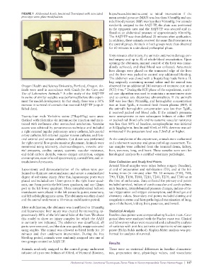
FIGURE 1  Abdominal Aortic Junctional Tourniquet with associated   https://www.biotimeinc.com) at initial intervention if the
              prototype torso plate modification.                mean arterial pressure (MAP) was less than 50mmHg and sys-
                                                                 tolic blood pressure (SBP) was less than 90mmHg. For animals
                                                                 randomly assigned to the AAJT-TP, the plate was positioned
                                                                 in the epigastric area and the AAJT-TP was secured and in-
                                                                 flated to an abdominal pressure of approximately 40mmHg.
                                                                 The AAJT-TP was then deflated 20 minutes after application.
                                                                 In addition, these animals received the same fluid treatment as
                                                                 the control group. Animals in both groups were then observed
                                                                 for 60 minutes in a simulated prehospital phase.
                                                                 Sixty minutes after injury, the animals underwent damage con-
                                                                 trol surgery and up to 3L of whole-blood resuscitation. Upon
                                                                 opening the abdomen, manual control of the liver was imme-
                                                                 diately achieved, and shed blood was collected. Atraumatic
                                                                 liver clamps were placed on the transected edge of the liver
                                                                 and the liver was packed to control any additional bleeding.
                                                                 The abdomen was closed with a Bogota bag made from a 1L
                                                                 bag originally containing normal saline and the animal was
              Oregon Health and Science University, Portland, Oregon. An-  observed for an additional 240 minutes of intensive care unit
                                                                         17
              imals were used in accordance with Guide for the Care and   (ICU) time.  During the ICU phase of the experiment, a criti-
                                    16
              Use of Laboratory Animals.  A pilot study of the AAJT-TP   cal care algorithm was used to maintain a normotensive state
              in swine of similar weights was performed before this experi-  and to correct any electrolyte abnormalities. If the animal’s
              ment for model development. In that study, there was a 50%   SBP was less than 90mmHg, and hemoglobin concentration
              increase in survival of animals that received AAJT-TP (unpub-  was at least 7g/dL, it received fresh frozen plasma (FFP). If
              lished data).                                      the animal’s hemoglobin concentration was less than 7g/dL,
                                                                 it  received  packed  red  blood cells.  If  the  animal’s pressures
              Twenty-four male Yorkshire swine (75kg–85kg) were anes-  were unresponsive to two subsequent  boluses of either FFP
              thetized with tiletamine via intramuscular injection and main-  or packed red blood cells and its systemic vascular resistance
              tained with isoflurane after orotracheal intubation. Vascular   was less than 80% of baseline, norepinephrine was initiated
              access was achieved by percutaneous technique and included   at 0.02μg/kg/min. Insulin and 50mL of 50% dextrose was ad-
              a right external jugular pulmonary artery catheter, left carotid   ministered if the potassium level was 5.5mEqL or higher.
              artery catheter, left external jugular venous catheter, and fem-
              oral arterial and venous catheters. Cut down was performed   At the completion of the experiment, animals were euthanized
              for right carotid flow-probe monitor placement. Animals were   and underwent necropsy and gross pathology assessment. Tis-
              monitored using telemetry, electrocardiogram, invasive arte-  sue samples were collected from the terminal ileum, kidney,
              rial  pressures,  cardiac  output,  systemic  vascular  resistance,   liver, pancreas, lung, and heart. These samples were sent for
              end-tidal carbon dioxide, venous oxygen saturation, oxygen   histological analysis by a certified veterinary pathologist.
              consumption, near-infrared spectroscopy, carotid flow, and in-
              traabdominal pressures.                            Data Collection and Study End Points
                                                                 Arterial blood samples were taken before surgery (baseline),
              Laparotomy and devascularization of the spleen were per-  at end of preparation and stabilization (T0), and at the fol-
              formed to eliminate autotransfusion and create a standardized   lowing times (in minutes) after T0: 30 minutes (T30), T60,
              degree of soft-tissue injury. After this, laparoscopic ports were   T90, T120, T150, T180, T210, T240, T270, and T300 or at
              placed and included two 12mm ports in the right lower quad-  the time of euthanasia. Data collected for primary end points
              rant, one 5mm port in the left lower quadrant, and one 12mm   included survival, indices of cardiovascular and cardiopulmo-
              port in the left lower quadrant. Three intraabdominal balloon   nary function, intraabdominal pressure changes, indices of tis-
              transducers were placed in the left paracolic gutter, mesentery,   sue oxygenation and oxygen consumption, and blood gas and
              and left subdiaphragmatic areas. The abdomen was then closed   chemistry values. Secondary end points included clotting and
              and the animal underwent a 10-minute stabilization period.  coagulation status and histopathological examination of lung,
                                                                 apex of the heart, kidney, liver, pancreas, and bowel.
              After stabilization, the abdomen was insufflated to 15mmHg
              and laparoscopic liver injury was created by transecting ap-  Statistical Analysis
              proximately 80% of the left lateral lobe of the liver. We chose   Baseline data points were compared using Student t test. Cate-
              this model to show an injury complex for which the AAJT   gorical data were analyzed with the Fischer exact test. Clinical
              is currently not indicated. The abdomen was desufflated, all   and laboratory values were measured and analyzed by analysis
              ports were removed, and the skin was quickly reapproximated   of variance with post hoc pairwise comparisons where appro-
              using staples. The animal was allowed to bleed freely for 10   priate (Holm-Sidak method). Kaplan-Meier analysis was per-
              minutes  and then  underwent  intervention.  During the  free   formed for analysis of survival.
              bleeding period, animals were randomly assigned into one of
              two groups: control or AAJT-TP.
                                                                 Results
              Animals randomly assigned to the control group underwent   There were no statistical differences in baseline characteris-
              infusion of up to two boluses of 500mL of Hextend (Biotime,   tics, preparation time, physiologic values, and viscoelastic

                                          Abdominal Aortic Junctional Tourniquet–Torso Plate in Noncompressible Torso Hemorrhage  |  107