
Page 78 - JSOM Summer 2018
P. 78
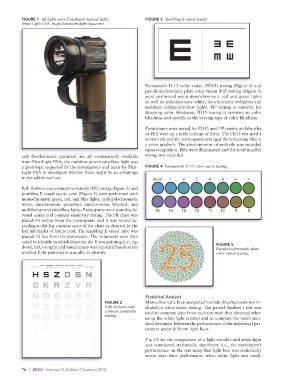
FIGURE 1  All lights were Tomahawk tactical lights    FIGURE 3  Tumbling E visual acuity.
          (First-Light USA, https://www.firstlight-usa.com).


                                                             Farnsworth D-15 color vision (FD15) testing (Figure 4) and
                                                             pseudoisochromatic plate color vision (PiP) testing (Figure 5)
                                                             were performed using monochromatic red and green lights
                                                             as well as polychromatic white, duochromatic red/green and
                                                             multihue red/green/yellow lights. PiP testing is sensitive for
                                                             detecting color blindness; FD15 testing is sensitive to color
                                                             blindness and specific to the varying type of color blindness.
                                                             Participants were seated for FD15 and PiP testing and the tiles
                                                             or PiPs were on a table in front of them. The FD15 test used a
                                                             control tile and the participants arranged the remaining tiles in
                                                             a color gradient. The absolute error of each tile was recorded
                                                             upon completion. PiPs were illuminated and the total number
          and duochromatic green/red are all commercially available   wrong was recorded.
          from First-Light USA; the multihue green/red/yellow light was
          a prototype requested by the investigators and made by First-  FIGURE 4  Farnsworth D-15 color vision testing.
          Light USA to investigate whether there might be an advantage
          to the additional hue.

          Pelli-Robson near contrast sensitivity (PR) testing (Figure 2) and
          tumbling E visual acuity cube (Figure 3) were performed with
          monochromatic green, red, and blue lights, ands polychromatic
          white, duochromatic green/red, duochromatic blue/red, and
          multihue green/red/yellow lights. Participants were standing for
          visual acuity and contrast sensitivity testing. The PR chart was
          placed 40 inches from the participants and it was scored ac-
          cording to the log contrast score of the chart as dictated by the
          last full triplet of letters read. The tumbling E vision cube was
          placed 10 feet from the participant. The volunteers were then
          asked to identify in which direction the E was pointing (i.e., up,
          down, left, or right) and visual acuity was recorded based on the               FIGURE 5
                                                                                          Pseudoisochromatic plate
          smallest E the participant was able to identify.                                color vision testing.


                                                             Statistical Analysis
                                            FIGURE 2         Monochromatic blue and polychromatic blue/red were not in-
                                            Pelli-Robson near   cluded in color-vision testing. The paired Student t test was
                                            contrast sensitivity   used to compare data from each test with that obtained when
                                            testing.
                                                             using the white light control and to compare the mean stan-
                                                             dard deviation between the performance of the individual par-
                                                             ticipant under different light hues.

                                                             P ≤ .05 for the comparison of a light variable and white light
                                                             was considered statistically significant (i.e., the participant’s
                                                             performance on the test using that light hue was statistically
                                                             worse than their performance when white light was used).


          76  |  JSOM   Volume 18, Edition 2/Summer 2018