
Page 142 - Journal of Special Operations Medicine - Fall 2017
P. 142
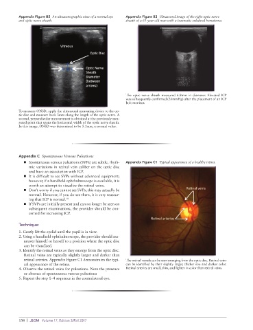
Appendix Figure B2  An ultrasonographic view of a normal eye   Appendix Figure B3  Ultrasound image of the right optic nerve
          and optic nerve sheath.                            sheath of a 61-year-old man with a traumatic subdural hematoma.


                                                             The optic nerve sheath measured 6.8mm in diameter. Elevated ICP
                                                             was subsequently confirmed (26mmHg) after the placement of an ICP
                                                             bolt monitor.
          To measure ONSD, apply the ultrasound measuring device to the op-
          tic disc and measure back 3mm along the length of the optic nerve. A
          second, perpendicular measurement is obtained at the previously mea-
          sured point that spans the horizontal width of the optic nerve sheath.
          In this image, ONSD was determined to be 5.1mm, a normal value.


          Appendix C  Spontaneous Venous Pulsations
            ■ ➤ Spontaneous venous pulsations (SVPs) are subtle, rhyth-  Appendix Figure C1  Typical appearance of a healthy retina.
               mic variations in retinal vein caliber on the optic disc
               and have an association with ICP.
            ■ ➤ It is difficult to see SVPs without advanced equipment;
               however, if a handheld ophthalmoscope is available, it is
               worth an attempt to visualize the retinal veins.
            ■ ➤ Don’t worry if you cannot see SVPs; this may actually be
               normal. However, if you do see them, it is very reassur-
               ing that ICP is normal. 10
            ■ ➤ If SVPs are initially present and can no longer be seen on
               subsequent examinations, the provider should be con-
               cerned for increasing ICP.


          Technique:
          1. Gently lift the eyelid until the pupil is in view.
          2. Using a handheld ophthalmoscope, the provider should ma-
            neuver himself or herself to a position where the optic disc
            can be visualized.
          3. Identify the retinal veins as they emerge from the optic disc.
            Retinal veins are typically slightly larger and darker than
            retinal arteries. Appendix Figure C1 demonstrates the typi-  The retinal vessels can be seen emerging from the optic disc. Retinal veins
            cal appearance of the retina.                    can be identified by their slightly larger, thicker size and darker color.
          4. Observe the retinal veins for pulsations. Note the presence   Retinal arteries are small, thin, and lighter in color than retinal veins.
            or absence of spontaneous venous pulsations
          5. Repeat the step 1–4 sequence in the contralateral eye.


          138  |  JSOM   Volume 17, Edition 3/Fall 2017