
Page 141 - Journal of Special Operations Medicine - Fall 2017
P. 141
• If patient is not responsive to voice, test central pain response (e.g., extensor posturing, flexor posturing,
and peripheral pain. withdrawal, localization).
➤ o Central pain: Apply a sternal rub or supraorbital NOTE: In an awake and cooperative patient, test-
pressure, and note the response (e.g., extensor pos- ing light touch is recommended. It is unnecessary to
turing, flexor posturing, localization). apply painful stimuli to an awake and cooperative
➤ o Peripheral pain: Apply nail bed pressure or take patient.
muscle between the fingers, compress, and rotate V. Gait
the wrist (do not pinch the skin). Muscle in the ax- • If the patient is able to walk, observe his/her casual
illary region and inner thigh is recommended. Ap- gait and note any instability, drift, sway, and so forth.
ply similar stimulus to all four limbs and note the
Appendix B Ultrasonographic Assessment of Optic Nerve Sheath Diameter
f a patient is unconscious (i.e., does not follow commands or ultrasonagraphic image of the optic nerve sheath can be
Iopen eyes spontaneously), they may have elevated ICP. There seen in Appendix Figure B2.
is no reliable test for elevated ICP available outside of a hos- 8. Once the optic nerve sheath is visualized, freeze the image
pital; however, optic nerve sheath diameter (ONSD) measure- on the screen.
ment is a rapid, safe, and easy-to-perform ultrasonographic 9. Using the device’s measuring tool, measure 3mm back
assessment that may help identify elevated ICP when more from the optic disc and then obtain a second measure-
definitive monitoring devices are not available. ment perpendicular to the first. The second measurement
should cover the horizontal width of the optic nerve
■ ➤ The optic nerve sheath directly communicates with the sheath (Appendix Figure B2). An abnormal ONSD is
intracranial subarachnoid space. Increased ICP, there- shown in Appendix Figure B3.
fore, displaces cerebrospinal fluid along this pathway. 10. Repeat the previous sequence in the opposite eye. Anno-
Normal ONSD is 4.1–5.9mm. 30 tate both ONSDs on the PFC Casualty Card.
–5
■ ➤ A 10 -MHz linear ultrasound probe can be used to ob- 11. ONSDs should be obtained, when possible, at regular in-
tain ONSDs. ONSD is measured from one side of the tervals to help assess changes in ICP, particularly when
optic nerve sheath to the other at a distance of 3mm the neurologic examination is poor and/or unreliable
behind the eye immediately below the sclera. 31 (i.e., with sedation). Serial measurements with progres-
■ ➤ In general, ONSDs >5.2mm should raise concern for sive diameter enlargement and/or asymmetry in ONSDs
clinically significant elevations in ICP in unconscious should be considered indicative of worsening intracranial
TBI patients. 5,32 The ONSD can vary significantly in hypertension.
normal individuals, so one single measurement may not CAUTION: ONSD measurements are contraindicated in globe
be helpful; however, repeated measurements that detect injuries. NEVER apply pressure to an injured globe.
gradual increases in ONSD over time may be more use-
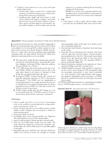
ful than a single measurement. Appendix Figure B1 Appropriate placement of the linear probe.
■ ➤ ONSD changes rapidly when the ICP changes, so it can
be measured frequently. If ONSD is used, it is best to
33
check hourly along with the neurologic examination.
Technique:
1. Check to make sure there is no eye injury. A penetrating
injury to the eyeball is a contraindication to ultrasound be-
cause it puts pressure on the eye.
2. Ensure the head and neck are in a midline position. Gentle
sedation and/or analgesia may be necessary to obtain ac-
curate measurements.
3. Ensure the eyelids are closed.
4. If available, place a thin, transparent film (e.g., Tegaderm;
3M, http://www.3m.com) over the closed eyelids.
5. Apply a small amount of ultrasound gel to closed eyelid.
6. Place the 10(–5)MHz linear probe over the eyelid. The
probe should be applied in a horizontal orientation (Ap-
pendix Figure B1) with as little pressure as possible applied
to the globe. Ultrasound gel is placed over a closed eyelid and the probe placed
horizontally over the eyelid, applying as little pressure to the globe
7. Manipulate the probe until the nerve and nerve sheath are as possible. If available, Tegaderm or other thin covering (e.g., Latex
visible at the bottom of screen. An example of a proper glove) should be placed over a closed eyelid for further protection.
Guideline: TBI Management in PFC | 137