
Page 92 - Journal of Special Operations Medicine - Spring 2017
P. 92
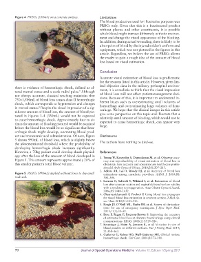
Figure 4  PRBCs (330mL) on a concrete surface.     Limitations
                                                             The blood product we used for illustrative purposes was
                                                             PRBCs only. Given that this is a fractionated product
                                                             without  plasma  and  other  constituents,  it  is  possible
                                                             whole blood might interact differently with the environ-
                                                             ment and change the visual appearance of the bleeding.
                                                             In addition, during actual wounding, there is likely to be
                                                             absorption of blood by the injured soldier’s uniform and
                                                             equipment, which was not pictured in the figures in this
                                                             article. Regardless, we believe the use of PRBCs allows
                                                             the reader to gain a rough idea of the amount of blood
                                                             loss based on visual estimation.


                                                             Conclusion
                                                             Accurate visual estimation of blood loss is problematic
                                                             for the reasons listed in this article. However, given lim-
                                                             ited objective data in the military prehospital environ-
          there is evidence of hemorrhagic shock, defined as al-
          tered mental status and a weak radial pulse.  Although   ment, it is unrealistic to think that the visual impression
                                                 9
          not  always  accurate,  classical  teaching  maintains  that   of blood loss will not affect patient-management deci-
          750–1,500mL of blood loss causes class II hemorrhagic   sions. Because of this, it is important to understand in-
          shock, which corresponds to hypotension and changes   herent biases such as overestimating small volumes of
          in mental status.  Despite the visual impression of a sig-  hemorrhage and overestimating large volumes of hem-
                        8
          nificant amount of blood loss, the amount of blood pic-  orrhage. We hope that the clinical images in this article
          tured in Figures 1–4 (330mL) would not be expected   give some perspective on this topic and illustrate that a
          to cause hemorrhagic shock. Approximately four to six   relatively small amount of bleeding, which would not be
          times the amount of bleeding pictured would be required   expected to cause hemorrhagic shock, can appear very
          before the blood loss would be so significant that hem-  large.
          orrhagic shock might develop, warranting blood prod-
          uct and tranexamic acid administration. Of note, Figure   Disclosures
          5 shows 990mL of blood loss, which is slightly below
          the aforementioned threshold where the probability of   The authors have nothing to disclose.
          developing hemorrhagic shock increases significantly.
          However, a 70kg patient could develop shock physiol-  References
          ogy after the loss of the amount of blood developed in   1. Yoong W, Karavolos S, Damodaram M, et al. Observer accu-
          Figure 5. This amount represents approximately 20% of   racy and reproducibility of visual estimation of blood loss in
          this smaller patient’s total blood volume.           obstetrics: how accurate and consistent are health-care profes-
                                                               sionals? Arch Gynecol Obstet. 2010;281:207–213.
                                                             2. Adkins AR, Lee D, Woody DJ, et al. Accuracy of blood loss
          Figure 5  PRBCs (990mL) applied without force to dry sand/  estimations among anesthesia providers.  AANA J. 2014;82:
          rock soil.                                           300–306.
                                                             3. Larsson C, Saltvedt S, Wiklund I, et al. Estimation of blood
                                                               loss after cesarean section and vaginal delivery has low validity
                                                               with a tendency to exaggeration. Acta Obstet Gynecol Scand.
                                                               2006;85:1448–1452.
                                                             4. Cheerranichanunth P, Poolnoi P. Using blood loss pictogram
                                                               for visual blood loss estimation in cesarean section. J Med As-
                                                               soc Thai. 2012;95:550–556.
                                                             5. Kragh JF, O’Neill ML, Beebe DF, et al. Survey of the indica-
                                                               tions for use of emergency tourniquets.  J Spec Oper Med.
                                                               2011;11(1):30–38.
                                                             6. Bose P, Regan F, Paterson-Brown S. Improving the accuracy
                                                               of estimated blood loss at obstetric haemorrhage using clinical
                                                               reconstructions. BJOG. 2006;113:919–924.
                                                             7. Kreutziger J, Haim  A, Jonsson K,  et al. Variation in size of
                                                               blood puddles on different surfaces. Eur J Emerg Med. 2014;
                                                               21:360–363.
                                                             8. Gutierrez G, Reines HD, Wulf-Gutierrez ME. Clinical review:
                                                               hemorrhagic shock. Crit Care. 2004;8:373–381.


          70                                      Journal of Special Operations Medicine  Volume 17, Edition 1/Spring 2017