Page 71 - Journal of Special Operations Medicine - Spring 2017
P. 71

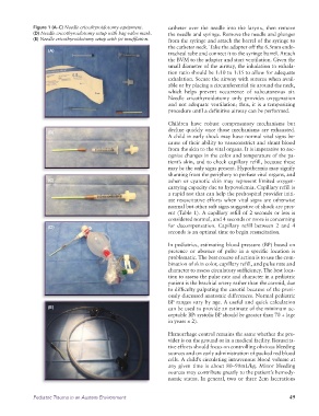
Figure 1 (A–C) Needle cricothyroidotomy equipment.    catheter over the needle into the larynx, then remove
              (D) Needle cricothyroidotomy setup with bag-valve mask.    the needle and syringe. Remove the needle and plunger
              (E) Needle cricothyroidotomy setup with jet insufflation.  from the syringe and attach the barrel of the syringe to
                                                                 the catheter neck. Take the adapter off the 6.5mm endo-
                   (A)
                                                                 tracheal tube and connect it to the syringe barrel. Attach
                                                                 the BVM to the adapter and start ventilation. Given the
                                                                 small diameter of the airway, the inhalation to exhala-
                                                                 tion ratio should be 1:10 to 1:15 to allow for adequate
                                                                 exhalation. Secure the airway with sutures when avail-
                                                                 able or by placing a circumferential tie around the neck,
                                                                 which  helps  prevent  occurrence  of  subcutaneous  air.
                                                                 Needle cricothyroidotomy only provides oxygenation
                                                                 and not adequate ventilation; thus, it is a temporizing
                                                                 procedure until a definitive airway can be performed.

                                                                 Children  have  robust  compensatory  mechanisms  but
                                                                 decline quickly once those mechanisms are exhausted.
                   (B)
                                                                 A child in early shock may have normal vital signs be-
                                                                 cause of their ability to vasoconstrict and shunt blood
                                                                 from the skin to the vital organs. It is imperative to rec-
                                                                 ognize changes in the color and temperature of the pa-
                                                                 tient’s skin, and to check capillary refill, because these
                                                                 may be the only signs present. Hypothermia may signify
                                                                 shunting from the periphery to perfuse vital organs, and
                   (C)                                           ashen or cyanotic  skin may represent  limited oxygen-
                                                                 carrying capacity due to hypovolemia. Capillary refill is
                                                                 a rapid test that can help the prehospital provider initi-
                                                                 ate resuscitative efforts when vital signs are otherwise
                                                                 normal but other soft signs suggestive of shock are pres-
                                                                 ent (Table 1). A capillary refill of 2 seconds or less is
                                                                 considered normal, and 4 seconds or more is concerning
                   (D)                                           for decompensation. Capillary refill between 2 and 4
                                                                 seconds is an optimal time to begin resuscitation.

                                                                 In pediatrics, estimating blood pressure (BP) based on
                                                                 presence or absence of pulse in a specific location is
                                                                 problematic. The best course of action is to use the com-
                                                                 bination of skin color, capillary refill, and pulse rate and
                                                                 character to assess circulatory sufficiency. The best loca-
                                                                 tion to assess the pulse rate and character in a pediatric
                                                                 patient is the brachial artery rather than the carotid, due
                                                                 to difficulty palpating the carotid because of the previ-
                                                                 ously discussed anatomic differences. Normal pediatric
                                                                 BP ranges vary by age. A useful and quick calculation
                   (E)                                           can be used to provide an estimate of the minimum ac-
                                                                 ceptable BP: systolic BP should be greater than 70 + (age
                                                                 in years × 2).

                                                                 Hemorrhage control remains the same whether the pro-
                                                                 vider is on the ground or in a medical facility. Resuscita-
                                                                 tive efforts should focus on controlling obvious bleeding
                                                                 sources and on early administration of packed red blood
                                                                 cells. A child’s circulating intravenous blood volume at
                                                                 any given time is about 80–90mL/kg. Minor bleeding
                                                                 sources may contribute greatly to the patient’s hemody-
                                                                 namic status. In general, two or three 2cm  lacerations


              Pediatric Trauma in an Austere Environment                                                      49
   66   67   68   69   70   71   72   73   74   75   76