
Page 26 - Journal of Special Operations Medicine - Spring 2017
P. 26
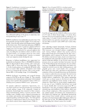
Figure 3  Establishment of arterial access and sheath   Figure 4  Out-of-hospital REBOA to facilitate patient
          placement for out-of-hospital REBOA.               salvage via resuscitation and damage control surgery. Arrow
                                                             highlights ER-REBOA catheter and 7F sheath in the right
                                                             common femoral artery.


                                                             Total blood required after REBOA inflation was a mean
          the calibrated marking on the device to determine opti-  of 3.75 units of whole blood (range, 2–6 units); three
          mal positioning in each case. 14,20                patients also received packed red blood cells (mean, 4.3
                                                             units; range, 1–8 units). Fresh frozen plasma (FFP) was
          REBOA insufflation was undertaken in aortic zone 1 for   administered to two patients: one received 1 unit of FFP
          three patients (average insertion depth, 47cm) and zone 3   and the other received 1 unit of FFP and 3 units of liq-
          (depth, 30cm) for the patient with fragmentation burden   uid plasma.
          to the lower torso. Total time from initiation of REBOA
          procedure (skin puncture or incision) to balloon inflation   After achieving surgical hemostasis, balloon deflation
          ranged from 5 to 8 minutes (Table 1). REBOA placement   was performed in a stepwise fashion over 3–5 minutes.
          and inflation were conducted by a general surgeon (two   This  maneuver was  tolerated  without complication  in
          cases, including the one instance where cut-down access   each instance. Balloon inflation time (aortic occlusion
          was performed) or an emergency physician (two cases).   time) ranged from 20 to 28 minutes for the three zone 1
          There  were no  fellowship-trained vascular  or trauma/  deployments, and 65 minutes for the zone 3 placement.
          acute care surgeons present during the care.       The balloon and femoral artery sheath were removed at
                                                             the completion of the procedure in each case before evac-
          Response to balloon insufflation was appreciated im-  uation to the next echelon of care. In two cases, manual
          mediately in all patients. Prior to ER-REBOA inflation,   pressure alone was used to facilitate hemostatic removal
          SBP was discernable in three patients, with a mean pre-  of the 7F sheath from the femoral artery. In each of these
          inflation SBP of 70mm Hg (range, 50–90mmHg; Table   instances, a handheld ultrasound device was used to ex-
          1). In the fourth patient, no measurable BP before infla-  amine the arterial access site after holding pressure, and
          tion was noted. After inflation, all patients were noted   palpable pulses were noted distally after completion.
          to have an immediate BP response, with a mean postin-  For a third case, there was a question on ultrasound ex-
          flation SBP of 118 mmHg (range, 110–120mmHg).      amination of femoral sheath hematoma and the decision
                                                             was made to explore the area and repair the arteriotomy
          REBOA  facilitated  resuscitation  and  surgical  damage   using interrupted polypropylene sutures; this was per-
          control of NCTH in all cases (Figure 4). Two patients   formed without complication. The patient undergoing
          underwent tube thoracostomy placement with minimal   initial open cut-down access to the femoral artery also
          output and no patient demonstrated evidence of thoracic   underwent uncomplicated suture closure of the arteri-
          bleeding above potential REBOA zone 1 placement.   otomy site. There were no access-related complications.
                                                             In one case of zone 1 REBOA, the balloon was noted
          All patients underwent exploratory laparotomy, dur-  to migrate distally after restoration of proximal BP to a
          ing which significant hemoperitoneum was encountered   hypertensive state that necessitated deflation, proximal
          in each instance (range, 1–3L estimated). Control of   repositioning, and reinflation of the balloon, which was
          mesenteric hemorrhage was required in three patients,   done without complication. After resuscitation and dam-
          along with intestinal, colonic, or gastric resection. Hem-  age control surgery, all patients were transported to a
          orrhage from a bladder source required intervention in   local facility for ongoing care. All patients survived the
          one patient and another with significant hepatic injury   approximate 2-hour transfer without incident and were
          required packing and hepatorraphy. The final patient   reported to be hemodynamically stable upon delivery to
          sustained a fragmentation injury to the external iliac ar-  the next level of care. No additional follow-up is avail-
          tery, which required direct surgical repair (Table 1).  able regarding their clinical course after transfer.


          4                                       Journal of Special Operations Medicine  Volume 17, Edition 1/Spring 2017