
Page 25 - Journal of Special Operations Medicine - Spring 2017
P. 25
the casualty collection point from the battlefield 10–15 Placement of the ER-REBOA catheter was undertaken
minutes away by local medics without formal training for all patients. Two patients underwent aortic balloon
or capabilities to implement tactical combat casualty occlusion before rapid sequence induction and intuba-
care guidelines. tion, and two had REBOA after rapid sequence induc-
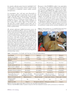
tion. Ultrasound was used to establish arterial access in
At presentation, clear vital signs were discernable in the common femoral artery in three patients (Figure 2).
three patients, with initial mean heart rate of 131 bpm For the fourth patient, the casualty with only a weakly
(range, 120–140 bpm), initial systolic blood pressure palpable carotid pulse on arrival, open femoral artery
(SBP) of 78mmHg (range, 70–90 mmHg), initial mean cut-down was performed. Access was achieved in the
respiratory rate of 24 breaths per minute (range, 24–25 common femoral artery in all four patients (Figure 3). A
breaths per minute), and initial oxygen saturation of 7F sheath was initially placed in each case, followed by
84% (range, 80% to 88%; Table 1). The fourth casu- an ER-REBOA catheter. Radiography was not available
alty had only a weakly palpable carotid pulse and no in this austere setting; thus, balloon catheter positioning
palpable femoral pulse, and demonstrated agonal respi- was performed using external anatomic landmarks and
ration at presentation.
Figure 2 Field-expedient use of a handheld ultrasound device
All patients underwent initial intravenous access via to facilitate location and cannulation for arterial access.
large-bore peripheral catheter, and one underwent ad-
ditional intraosseous line placement before the initiation
of surgical intervention. All patients received an initial
2 units of whole blood immediately after establishment
of venous access and received antibiotics and 1g of
tranexamic acid (TXA) during early resuscitation. Fo-
cused abdominal sonography for trauma (FAST) exami-
nation using the ultrasound Vscan device (V-Scan; GE
Healthcare, http://www3.gehealthcare.com/) was under-
taken before operation for three of the patients, two of
whom demonstrated significant hemoperitoneum and
one whose FAST scan showed blood in the pelvis. It was
determined that the remaining patient needed emergent
exploratory laparotomy, based on the wounding pattern
and physiologic status.
Table 1 Demographics, Use Details, and Outcomes of Out-of-Hospital REBOA
Case Details Patient 1 Patient 2 Patient 3 Patient 4
Mechanism of injury Gunshot Gunshot Gunshot Explosive
SBP before REBOA 90mmHg 70mmHg 50mmHg Unmeasurable
inflation
Means of femoral Percutaneous Percutaneous Percutaneous Cut down
artery access
Depth (cm)/intended zone 48cm/zone 1 47cm/zone 1 45cm/zone 1 30cm/zone 3
Time to aortic occlusion 5 minutes 5 minutes 7 minutes 8 minutes
SBP after REBOA inflation* 120mmHg 120mmHg 110mmHg 120mmHg
Duration of aortic occlusion 28 minutes 20 minutes 27 minutes 65 minutes
Operation after REBOA Laparotomy Laparotomy Laparotomy Laparotomy
Surgical/bleeding source Mesenteric/bladder Mesenteric bleeding Mesenteric/hepatic External iliac
control interventions bleeding source control source control bleeding source control artery repair
Method of sheath removal Manual pressure Manual pressure Open repair of artery Open repair of artery
Catheter-related No No No No
complication
REBOA provider Surgeon Emergency medicine Emergency medicine Surgeon
REBOA, resuscitative endovascular balloon occlusion of the aorta; SBP, systolic blood pressure.
*Inflation of the ER-REBOA catheter occurred in conjunction with other resuscitative maneuvers such as administration of whole blood.
REBOA in CCC Setting 3