
Page 126 - Journal of Special Operations Medicine - Spring 2016
P. 126
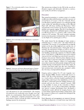
Figure 1  Photo indicating width of area of fluctuance on   The patient was evaluated in the ED by the on-call or-
          patient’s lower back.                              thopedic surgeon  and ultimately referred  to a tertiary
                                                             care facility for definitive management.

                                                             Discussion

                                                             The typical presentation is a patient report of a tender,
                                                             swollen area often with fluctuance and either an acute or
                                                             remote history of trauma to the site. Clinically, Morel-
                                                             Lavallée lesions typically present as an enlarging painful
                                                             lesion within the anterolateral portion of the affected
                                                                                                      2
                                                             thigh with soft tissue swelling and fluctuance.  Other
                                                             reported sites of involvement are the trunk, lumbar, pre-
                                                             patellar, and scapular regions. The 2012 Case Reports in
                                                             Orthopedics described 29 published reports of Morel-
                                                             Lavallée lesions from 14 countries, with a total of 204
                                                                                  3
                                                             lesions in 195 patients.  The most common anatomic
                                                             location was the greater trochanter/hip (36%), followed
                                                             by the thigh (24%) and the pelvis (19%). 3
          Figure 2  Photo indicating of area of fluctuance on patient’s
          lower back.                                        The cavity formed by the shearing of tissue creates a
                                                             potential space or “sixth cavity” into which blood can
                                                             accumulate. The amount of accumulated blood depends
                                                             mainly on the size of the degloved area and the rate of
                                                             bleeding into this potential space. During the initial as-
                                                             sessment, the mechanism of injury, as well as any large
                                                             area of bruising in the absence of an obvious source of se-
                                                             vere bleeding, should raise suspicion for Morel-Lavallée
                                                             lesion.  Large Morel-Lavallée lesions may require surgical
                                                                  4
                                                             interventions for both bleeding control and to prevent in-
                                                                                 5
                                                             fection or skin necrosis. However, these effusions may be
                                                             initially missed or may take some time to develop, which
                                                             can make clinical diagnosis difficult. In longstanding

                                                             cases, these lesions may subsequently enlarge and become
                                                             painful, leading to misdiagnosis of soft tissue tumor. In
                                                             contrast, these lesion types have also been described to
          Figure 3  Transverse soft tissue ultrasound image of lumbar   have spontaneously decreased in size, remained a stable
          area, demonstrating deep, fascial anechoic fluid collection.  size over time, or progressively enlarged. 1
                                                             Imaging options include US, CT, and magnetic reso-
                                                             nance imaging (MRI). Features noted with US are re-
                                                             lated  to the age  of the  hematoma,  which will  appear
                                                             as a focal complex collection located superficial to the
                                                             muscle plane and deep to the hypodermis. A CT scan
                                                             will show a fluid level from the settling of blood com-
                                                             ponents and can also show a capsule if present. MRI
                                                             is the modality of choice in the evaluation of Morel-
                                                             Lavallée lesion, as it is able to discriminate chronicity
                                                             and internal contents. Radiologists have developed a

                                                             classification system defined by imaging features such
          and subcutaneous fat were unremarkable. The imaging   as lesion shape, signal characteristics, enhancement, and
          characteristics and mechanism were thought most sug-  the presence or absence of a capsule that distinguish the
          gestive of a posttraumatic Morel-Lavallée fluid collec-  different types. 6,7
          tion. A subsequent computed tomography (CT) scan
          performed to further evaluate the lesion confirmed the   Early diagnosis and management are essential, because
          findings reported by ultrasound.                   any delay in diagnosis or missed lesion may lead to the


          110                                     Journal of Special Operations Medicine  Volume 16, Edition 1/Spring 2016