
Page 85 - Journal of Special Operations Medicine - Spring 2015
P. 85
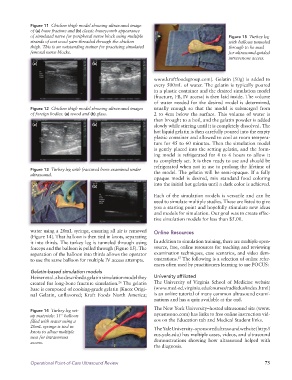
Figure 11 Chicken thigh model showing ultrasound image
of (a) bone fracture and (b) classic honeycomb appearance
of simulated nerve for peripheral nerve block using multiple Figure 15 Turkey leg
strands of wet wool yarn threaded through the chicken with balloon tunneled
thigh. This is an outstanding trainer for practicing simulated through to be used
femoral nerve blocks. for ultrasound-guided
intravenous access.
(a) (b)
www.kraftfoodsgroup.com). Gelatin (50g) is added to
every 500mL of water. The gelatin is typically poured
in a plastic container and the desired simulation model
(fracture, FB, IV access) is then laid inside. The volume
of water needed for the desired model is determined,
Figure 12 Chicken thigh model showing ultrasound images usually enough so that the model is submerged from
of foreign bodies: (a) wood and (b) glass. 2 to 4cm below the surface. This volume of water is
then brought to a boil, and the gelatin powder is added
(a) (b) slowly while stirring until it is completely dissolved. The
hot liquid gelatin is then carefully poured into the empty
plastic container and allowed to cool at room tempera
ture for 45 to 60 minutes. Then the simulation model
is gently placed into the setting gelatin, and the form
ing model is refrigerated for 4 to 6 hours to allow it
to completely set. It is then ready to use and should be
refrigerated when not in use to prolong the lifetime of
Figure 13 Turkey leg with fractured bone examined under the model. The gelatin will be semiopaque. If a fully
ultrasound.
opaque model is desired, mix standard food coloring
(a) (b) into the initial hot gelatin until a dark color is achieved.
Each of the simulation models is versatile and can be
used to simulate multiple studies. These are listed to give
you a starting point and hopefully stimulate new ideas
and models for simulation. Our goal was to create effec
tive simulation models for less than $5.00.
water using a 20mL syringe, ensuring all air is removed Online Resources
(Figure 14). That balloon is then tied in knots, separating
it into thirds. The turkey leg is tunneled through using In addition to simulation training, there are multiple open
forceps and the balloon is pulled through (Figure 15). The source, free, online resources for teaching and reviewing
separation of the balloon into thirds allows the operator examination techniques, case scenarios, and video dem
27
to use the same balloon for multiple IV access attempts. onstrations. The following is a selection of online refer
ences often used by practitioners learning to use POCUS:
Gelatin-based simulation models
Heiner et al. also described a gelatin simulation model they University affiliated
created for longbone fracture simulation. The gelatin The University of Virginia School of Medicine website
26
base is composed of cookinggrade gelatin (Knox Origi (www.meded.virginia.edu/courses/rad/edus/index.html)
nal Gelatin, unflavored; Kraft Foods North America; is an online tutorial of many common ultrasound exami
nations and has a quiz available at the end.
The New York University–hosted ultrasound site (www.
Figure 14 Turkey leg set-
up materials: 11" balloon nyuemsono.com) has links to free online instruction vid
filled with water using a eos on the Education tab and Medical Student links.
20mL syringe is tied in The Yale University–sponsored ultrasound website (http://
knots to allow multiple eus.yale.edu) has multiple cases, videos, and ultrasound
uses for intravenous
access. demonstrations showing how ultrasound helped with
the diagnosis.
Operational Point-of-Care Ultrasound Review 75