
Page 84 - Journal of Special Operations Medicine - Spring 2015
P. 84
®
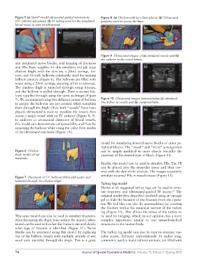
Figure 5 (a) Spam model ultrasound-guided intravenous Figure 8 (a) Chicken with two lines placed. (b) Ultrasound
(IV) catheter placement. (b) IV tubing used for the simulated guidance used to access the lines.
blood vessel as seen on ultrasound.
(a) (b)
(a)
(b)
Figure 9 Ultrasound images of (a) simulated vessels and (b)
the catheter in the vessel lumen.
and peripheral nerve blocks, and imaging of fractures
and FBs. Basic supplies for this simulator include large (a) (b)
chicken thighs with the skin on, a 20mL syringe, for
ceps, and 11inch balloons commonly used for making
balloon animals (Figure 6). The balloons are filled with
water using a 20mL syringe, ensuring all air is removed.
The chicken thigh is tunneled through using forceps,
and the balloon is pulled through. Then a second bal
loon is pulled through using the same technique (Figure
7). We recommend using two different colors of balloon Figure 10 Ultrasound images demonstrating (a) simulated
to ensure the balloons are not crossed when tunneling blood flow in vessels and (b) compressibility.
them through the thigh. Once both “vessels” have been (b)
placed, ultrasound is used to visualize the vessels then (a)
access a single vessel with an IV catheter (Figure 8, 9).
In addition to ultrasound detection of blood vessels,
this model can demonstrate compressibility and flow by
squeezing the balloon while using the color flow modes
of the ultrasound machines (Figure 10).
model for simulating femoral nerve blocks or other pe
ripheral blocks. The “vessel” and “nerve” arrangement
Figure 6 Chicken can be simply modified to more closely resemble the
thigh model set-up anatomy of the desired type of block (Figure 11).
materials.
Finally, this model can be used to simulate FBs. The FB
can be placed into the muscular tissue and then cov
ered with the skin of the chicken. The images accurately
simulate retained FBs in muscle tissue (Figure 12).
Figure 7 Placement of 11" balloons filled with water and
tunneled through the chicken thigh.
Turkey leg model
Heiner et al. suggested turkey legs can be used to simu
late fractures and ultrasoundguided IV access. The
25
original model they described involved using an opaque
gel to hide the location of the fracture from the opera
tor. We feel this can also be accomplished by creating
the fracture within the muscular section of the turkey
leg (Figure 13). This allows the tissue of the turkey to
This same model can also be used to simulate fractures. be used for imaging, which, in our opinion, has a more
After fracturing the thigh bone within the muscle, ultra complex appearance similar to true musculoskeletal
sound can be used to localize the fracture site and clarify structures in the human body.
what type of fracture is identified (Figure 11). Nerve
blocks can be simulated using this model by replacing The turkey leg model can also be used to simulate vas
one of the balloon vessels with multiple strands of wet cular access. Balloons approximately 11 inches long,
wool yarn tunneled through the thigh. This is a great commonly used to make balloon animals, are filled with
74 Journal of Special Operations Medicine Volume 15, Edition 1/Spring 2015