
Page 83 - Journal of Special Operations Medicine - Spring 2015
P. 83
®
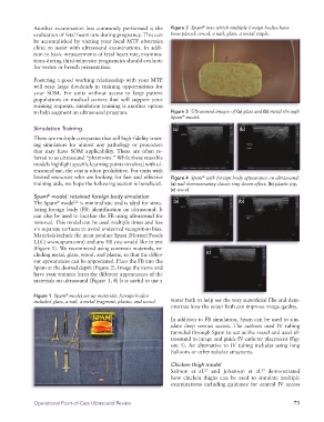
Another examination less commonly performed is the Figure 2 Spam into which multiple foreign bodies have
evaluation of fetal heart rate during pregnancy. This can been placed: wood, a nail, glass, a metal staple.
be accomplished by visiting your local MTF obstetrics
clinic to assist with ultrasound examinations. In addi
tion to basic measurements of fetal heart rate, examina
tions during thirdtrimester pregnancies should evaluate
for vertex or breech presentation.
Fostering a good working relationship with your MTF
will reap large dividends in training opportunities for
your SOM. For units without access to large patient
populations or medical centers that will support your
training requests, simulation training is another option
to help augment an ultrasound program. Figure 3 Ultrasound images of (a) glass and (b) metal through
Spam model.
®
Simulation Training (a) (b)
There are multiple companies that sell highfidelity train
ing simulators for almost any pathology or procedure
that may have SOM applicability. These are often re
ferred to as ultrasound “phantoms.” While these reusable
models highlight specific learning points involved with ul
trasound use, the cost is often prohibitive. For units with
limited resources who are looking for fast and effective Figure 4 Spam with foreign body appearance on ultrasound:
®
training aids, we hope the following section is beneficial. (a) nail demonstrating classic ring-down effect, (b) plastic pig,
(c) wood
Spam model: retained foreign body simulation
®
The Spam model is minimalistic and is ideal for simu (a) (b)
22
®
lating foreign body (FB) identification on ultrasound. It
can also be used to localize the FB using ultrasound for
removal. This model can be used multiple times and has
six separate surfaces to avoid undesired recognition bias.
Materials include the meat product Spam (Hormel Foods
LLC; www.spam.com) and any FB you would like to test
(Figure 1). We recommend using common materials, in
cluding metal, glass, wood, and plastic, so that the differ (c)
ent appearances can be appreciated. Place the FB into the
Spam at the desired depth (Figure 2). Image the items and
have your trainees learn the different appearances of the
materials on ultrasound (Figure 3, 4) It is useful to use a
Figure 1 Spam model set-up materials: foreign bodies
®
included glass, a nail, a metal fragment, plastic, and wood. water bath to help see the very superficial FBs and dem
onstrate how the water bath can improve image quality.
In addition to FB simulation, Spam can be used to sim
ulate deep venous access. The authors used IV tubing
tunneled through Spam to act as the vessel and used ul
trasound to image and guide IV catheter placement (Fig
ure 5). An alternative to IV tubing includes using long
balloons or other tubular structures.
Chicken thigh model
Salmon et al. and Johanson et al. demonstrated
23
24
how chicken thighs can be used to simulate multiple
examinations including guidance for central IV access
Operational Point-of-Care Ultrasound Review 73