
Page 138 - Journal of Special Operations Medicine - Spring 2015
P. 138
Cutaneous Leishmaniasis
Mark W. Burnett, MD
ABSTRACT
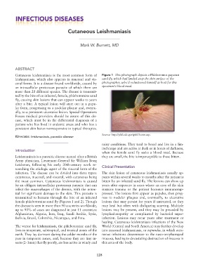
Cutaneous leishmaniasis is the most common form of Figure 1 This photograph depicts a Phlebotomus papatasi
leishmaniasis, which also appears in mucosal and vis sand fly, which had landed atop the skin surface of the
ceral forms. It is a disease found worldwide, caused by photographer, who’d volunteered himself as host for this
an intracellular protozoan parasite of which there are specimen’s blood meal.
more than 20 different species. The disease is transmit
ted by the bite of an infected, female, phlebotomine sand
fly, causing skin lesions that can appear weeks to years
after a bite. A typical lesion will start out in a papu
lar form, progressing to a nodular plaque and, eventu
ally, to a persistent ulcerative lesion. Special Operations
Forces medical providers should be aware of this dis
ease, which must be in the differential diagnosis of a
patient who has lived in endemic areas and who has a
persistent skin lesion nonresponsive to typical therapies.
Source: http://phil.cdc.gov/phil/home.asp.
Keywords: leishmaniasis, parasitic disease
rainy conditions. They tend to breed and live in a lim
ited range and are active at dusk or in hours of darkness,
Introduction
when the female sand fly seeks a blood meal. Because
Leishmaniasis is a parasitic disease named after a British they are small, the bite is imperceptible to those bitten.
Army physician, LieutenantGeneral Sir William Boog
Leishman, following his early 20thcentury work re Clinical Presentation
searching the etiologic agent of the visceral form of the
infection. The disease can be divided into three types: The skin lesion of cutaneous leishmaniasis usually ap
cutaneous, mucosal, and visceral, with cutaneous being pears within several weeks to months after the patient is
the most common. Cutaneous leishmaniasis is caused bitten by an infected sand fly. The lesions can show up
by an obligate intracellular protozoan parasite that can years after exposure in cases where an area of the skin
infect the macrophages of the dermis, with the poten sustains trauma or the patient becomes immunosup
tial for significant damage to the skin. This parasite is pressed. The lesions first appear as papules, then prog
transmitted to humans through the bite of an infected ress to nodular plaques and, eventually, to ulcerative
female phlebotomine sand fly (Figures 1 and 2). Though lesions that may persist for years if untreated, or they
the disease is seen in more than 90 countries worldwide, may heal but often with disfiguring scarring. Multiple
up to 90% of cases are diagnosed in just 11 countries: lesions may be present, and they may be preceded by
Afghanistan, Algeria, Iran, Iraq, Saudi Arabia, Syria, lymphadenopathy or complicated by bacterial super
Bolivia, Brazil, Colombia, Nicaragua, and Peru. infection. Lesions may recur years after treatment or
healing. Cutaneous leishmaniasis infections of the New
The vector for leishmaniasis, the phlebotomine sand fly, World (Central and South America) may further develop
lives in temperate, subtropical, and tropical zones of the into mucosal leishmaniasis, or espundia, in which cuta
earth. They lay dormant during the colder months of the neous infections disseminate to the nasooropharngeal
year in temperate zones, and, because they are tiny in mucosa, leading to devastating destruction of mucosa in
sects (2–3mm) that fly poorly, are less active in windy and this area of the body.
128