
Page 11 - Journal of Special Operations Medicine - Spring 2014
P. 11
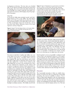
development of infection. The burn sites were checked   Figure 5  Case 2: Debridement was performed on the blister
              after 24 and 48 hours through the temporary removal of   necrotic contours on both arms, and accurate cleansing
              the bandage; due to the use of sterile saline infusion, the   and washing were performed. Both upper limbs were then
              same bandage was reused. Using the same gauze ban-  wrapped with moist silver-nylon dressing rolls (15cm × 165cm)
              dage will save resources while keeping intact the proper-  in direct contact with the entire wounded skin surface.
                                                                 On both hands we used silver-nylon gloves (size XL).
              ties of the gauze.                                 Subsequently, silver-nylon dressing was again covered with
                                                                 saline-moistened Kerlix bandage to maintain moisture.
              Case Two
              A 22-year-old white man sustained second- and third-
              degree burns on 27% of his TBSA. The third-degree
              burns were to the upper limbs showing skin area of red-
              ness and broken blisters with radial pulse at the extrem-
              ity. The second-degree burns were on the face, and his
              nose hairs were burnt, indicating a burn in the upper
              respiratory airways (Figure 4).

              Figure 4  Case 2: The third-degree burns on the upper limbs
              with radial pulse at the extremity showing skin area of
              redness and broken blisters.


                                                                 intensive care unit for 48 hours until arrival of the medi-
                                                                 cal evacuation team, which transported the patient to
                                                                 the combat support hospital in south Afghanistan and
                                                                 then to Germany. (In both patients, the cause of delay
                                                                 in transfer was a thunderstorm.) In these 48 hours, no
                                                                 dressing changes were made. The dressings were kept
                                                                 moist by spraying saline every 5 to 6 hours. The pa-
                                                                 tient remained stable. At dressing check after 24 and 48
                                                                 hours, no anomalies or infection was evident, and the
                                                                 silver-nylon dressing was not adhering to the wound.

              On arrival at the Role 2 facility, the Soldier was con-  Like for patient 1, it was not possible to perform wound
              scious with a GCS score of 13, and he had no fractures   cultures due to the logistical conditions of the operat-
              or internal organ lesions. Immediate ATLS protocols   ing environment, nor was it possible to follow up in the
              were applied. We opted for intraosseous access at the   subsequent days due to the quick transfer of the patient.
              tibia due to difficulties in obtaining venous access. This   Again, however, the bacteriostatic activity of the Silver
              was later replaced by a central venous catheter in the   Nylon dressing was evident in the first 48 hours because
              right subclavian vein once in the operating room. There   there was no clinical development of infection. And the
              he was intubated for protection of his airways and   burn sites were checked after 24 and 48 hours through
              avoidance of respiratory distress. Fluid resuscitation ac-  the temporary removal of the bandage; due to the use
              cording to the Parkland formula was initiated as well   of sterile saline infusion, the same bandage was reused.
              as intraosseous antibiotic therapy. Debridement of the
              burn was performed on both arms, and cleansing and
              washing were performed. Both upper limbs were then   Discussion
              wrapped up with moist silver-nylon dressing rolls (15cm   The bactericidal properties of silver are widely docu-
              × 165cm) in direct contact with the entire skin wound   mented. For silver ions (Ag ) to exhibit their bactericidal
                                                                                        +
              surface. Silver-nylon gloves (size XL) were used on both   properties, they must be available in solution. The ef-
              hands. Application of the gloves was easy due to their   ficacy of their action depends on the ion concentration
              elastic properties and did not cause any unintended fric-  on the bacterial surface. Silver ions act instantly, altering
              tion with the burn wound (Figure 5). The silver-nylon   bacteria cell walls, blocking the enzymatic respiratory
              dressing was again covered with saline-moistened gauze   system, and damaging microbial DNA. Fortunately, sil-
              bandage to maintain moisture. The Soldier stayed in the   ver ions are not toxic for human cells in vivo. The only


              Silver-Nylon Dressing to Treat Combat Burns in Afghanistan                                       3