


Page 92 - 2026 SMOG
P. 92
MWD NORMAL PARAMETERS

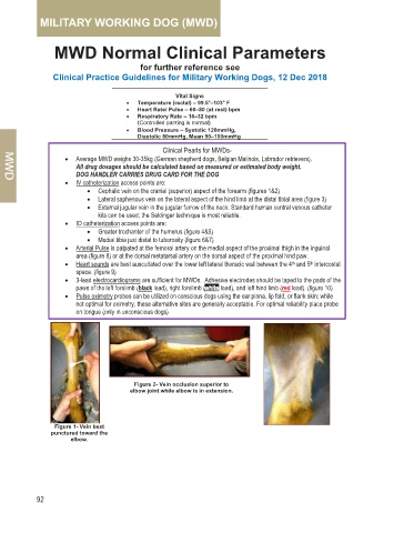
                                              Figure 2  Occlusion of
                                              cephalic vein.


                                    IVC NOTE:
                                    •  Use the cephalic or lateral
                                     saphenous vein for routine blood
                                     sampling, drug administration, and
                                     IV fluid therapy
                                    •  Use the external jugular vein for
                                     long-term IV fluid therapy, large-
                                     volume fluid delivery, and repeated
                                     blood sampling
                                    •  To access the external jugular vein,   Figure 4  Lateral
                                     standard human central venous   saphenous vein
         Figure 1  Cephalic   Figure 3  Cephalic IV   catheter kits can be used; the   location.
         location.     catheter placement.
                                     Seldinger technique is most reliable.
                               (left) Figure 5  Landmark
                               for IO catheterization on the
                               upper medial aspect of the
                               hind leg
                                  (right) Figure 6  Location
                                 for palpation of the femoral
                                      arterial pulse while
                                       dog is standing.
                             (left) Figure 7  Placement
                             of ECG electrode pad.


                                 (right) Figure 8  Placement
                                of human pulse oximeter, finger
                                       probe on tongue.
                    ECG NOTE                   PULSE OXIMETRY NOTE
         •  Canine ECG complexes resemble human   •  Human pulse oximetry probes (typically finger
           complexes with minor variations in key ECG   probes) are best placed on the tongue for
           intervals and possible inverted T-waves  optimal reliability in unconscious, sedated or
         •  Electrode or ultrasound gel may be required   anesthetized MWDs but can also be placed on
           between the electrode and footpads to improve   the prepuce or vulva.
           contact                       •  In conscious dogs, use the ear pinna, lip fold,
                                           or flank skin fold. Although these are not
                                           optimal for pulse oximetry, these alternate sites
                                           generally yield reliable results.


        84  •   MILITARY WORKING DOG (MWD)

