
Page 58 - 2022 Ranger Medic Handbook
P. 58
Eye Injury
Penetrating injuries to eye globe or fracture of the orbit must be assessed with any facial trauma in the combat setting.
In the combat setting, penetrating wounds of the eye may be very common from shrapnel and debris. Blunt trauma that
may disrupt the integrity of the globe may be seen during facial trauma from falls, PLF, FRIES landings, hand-to-hand
combat, or MVA-type collisions. The primary management in any setting includes a rigid eye shield that does not put
pressure on the globe of the eye. Avoid any manipulation of eye or eye globe if penetrating injury is suspected. Infec-
SECTION 2 post-traumatic endophthalmitis.
tion may cause later permanent loss of vision, so early broad-spectrum systemic antibiotic therapy is critical to prevent
TCCC Application
Care Under Fire: Stop life-threatening bleeding.
Tactical Field Care/Tactical Evacuation: If a penetrating eye injury is noted or suspected: Perform a rapid field test of
visual acuity and document findings. Cover the eye with a rigid eye shield (NOT a pressure patch). Give ondansetron
4–8mg IV/IM/ODT/PO to prevent vomiting and the subsequent increase in IOP. Ensure that the 400mg moxifloxacin
tablet in the combat pill pack is taken if possible. If able to take PO: moxifloxacin, 400mg PO once a day. If unable to
take PO: ertapenem, 1g IV/IM once a day.
Extended Care
Retrobulbar Hematoma: Blunt or penetrating periocular trauma may result in orbital bleeding. As the pressure in the
orbital compartment is progressively elevated, the intraocular pressure will also rise. If intraocular pressure rises to a
sufficiently high level, either central retinal artery occlusion or damage to the optic nerve may ensue and vision may be
permanently lost in the eye. Signs and symptoms of retrobulbar hemorrhage include pain, periorbital ecchymosis, pro-
gressive proptosis (bulging forward of the eye), decreased vision, diffuse subconjunctival hemorrhage, and an afferent
papillary defect. The definitive management for this disorder is a lateral canthotomy.
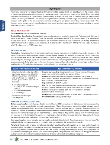
Rapid Field Visual Acuity Test Eye Examination (TRAUMA)
Visual acuity is the vital sign of the Inspect surrounding structures: Inspect the symmetry of the eyes,
eye in your assessment. Vision in eyebrows, and orbital area for any abnormalities.
affected eye should be checked Eyelids: Inspect the patient’s lightly closed eyelids for symmetry,
with unaffected eye closed. A fasciculation, tremors, and presence of eyelashes. While closed, look to
simple quantification is from best ensure eyelids close completely.
to worst:
Pupils: PERRLA, distortion, size
1. Able to read print Iris: Details clear, blood in anterior chamber, evidence of iris tissue in
2. Can count the number of fingers cornea or limbus, laceration or indication of penetrating trauma
held up
3. Can see hand motion Sclera: Obvious lacerations, dark iris or uveal tissue, redness,
4. Can see light subconjunctival hemorrhage
Cornea: Obvious defects (laceration or penetration), iris tissue in cornea
Document the finding on
Casualty Card. Ocular motion: Inability to move eye
Standard Visual Acuity Test
Distant visual acuity is tested using a Snellen chart with patient 20 ft away in a well-lighted area. Test each separately,
with one eye being covered while testing the opposite eye. Allow a few moments for eyes to adjust between tests. If
patient wears corrective vision, record two separate tests: one with and one without correction. Documentation is re-
corded as a fraction in which the numerator indicates the distance from the chart (20), and the denominator indicates the
distance at which the average eye can read the line (i.e., 20/40 indicates the patient is reading at 20 ft what the average
eye can read at 40 ft). Tell patient to read the line most clear to them and then proceed to the next distance level. Record
the distance in which the patient can still accurately read the text.
Near visual acuity is tested using the same principles as distant vision, but with Rosenbaum pocket vision screener. The
patient holds the card approximately 14 inches from the eye and reads the smallest line possible.
Peripheral visual acuity is tested using the confrontation test. Stand facing the patient at eye level and test each eye
separately. While the patient covers one eye, you cover the opposing eye (patient, left; examiner, right). Fully extend your
arm midway between yourself and the patient and then move it centrally with the fingers moving. Have the patient tell
you when the moving fingers are first seen. Compare the patient’s response with your response in the upper, lower, left,
and right spectra. Record as the estimated degrees of vision, with directly ahead being 0 degrees.
44 SECTION 2 PRIMARY TRAUMA PROTOCOLS