
Page 99 - JSOM Fall 2025
P. 99
to the standard Doppler ultrasonography measurements. Ob- Procedure
taining and scoring two-dimensional ultrasound recordings is Two test stations were set up; each attended by a vascular
easier to master due to its visual nature. Recent developments, surgeon and an assistant. The participants entered the test
4
such as ultrasound probe connectivity to smartphones and stations in randomly assigned buddy pairs and received in-
tablets, have made ultrasound devices portable and therefore structions regarding the study protocol. The aforementioned
more usable in operational settings outside of hospitals, such pairs performed the measurements on each other.
as on the waterside. Given the strategic importance of target-
ing critical infrastructure in underwater operations, exploring First, the performing participant was allowed 2 minutes of
innovative approaches to enhance operational readiness could practice time with the abdominal ultrasound probe on their
significantly strengthen the competitive advantage of maritime buddy, receiving feedback from the instructor. The participant
special operations forces (SOF) units. was then asked to remove the probe from the patient and start
the official procedure. The buddy was positioned on the ex-
Submitted data obtained by our study group suggests that amination bench in the supine position. The performer was
bubble scores of the inferior vena cava (IVC) and popliteal allowed a maximum of 2 minutes to obtain a 5-second video
vein (PV) show a significant correlation with the bubble-load of the IVC. Subsequently, the patient stood up, and the per-
of the diver, as determined by Doppler analysis. Thus, we hy- former was again allowed a maximum of 2 minutes to obtain
pothesized that with the accumulation of additional scientific a 5-second video of the PV. The performers were tasked with
evidence, vascular ultrasonography of these vessels may be- identifying the correct vessel themselves and did not receive
come an alternative method for detection of elevated decom- any instructions regarding identification during the procedure.
pression stress levels in the future. This could allow military Once the performers stated that they felt confident that they
(SOF) divers to become less dependent on scarce Doppler had identified the correct vessel, they recorded and saved the
analysis experts and enhance operational readiness. 5-second video.
The first step in evaluating the viability of self-monitoring of Scoring
decompression stress, and possibly on-site titration of dive Three outcomes were measured in this study: (1) observer
profiles, performed by dive teams on the waterside involves assessment of the performance, (2) self-perceived procedure
assessing the capacity of military combat medics to acquire experience (supplemental Figure 1), and (3) video recording
proficiency in generating high-quality vascular ultrasound quality. Performers were assessed by a vascular surgeon on
recordings. their preparation of the procedure, knowledge of materials
and instruments, time and motion, the progression of the pro-
The primary aim of this study was to assess the feasibility cedure and forward planning, their ability to adapt to individ-
of training combat medics to perform ultrasound measure- ual anatomical circumstances, and their overall performance
ments of the IVC and the PV with the use of a microteaching (Supplemental Table 1); adapted from the Objective Struc-
program. The secondary aim was to evaluate the quality of tured Assessment of Technical Skills [OSATS]). 5
two-dimensional recordings made by combat medics.
Participants were asked to fill in a scoring sheet about their
self-perceived experience, visibility of the vessels, and opinion
Methods
on performing vascular ultrasound on the waterside. Scoring
Participants options ranged from 1 (strongly disagree/very challenging) to
The group consisted of 26 SOF combat medics, some of whom 5 (strongly agree/very easy) (Appendix 1).
were also SOF divers. Five participants were certified NATO
Special Operations Combat Medics (NSOCMs). An NSOCM Recording quality was scored by a blinded vascular surgeon and
is a servicemember who provides TCCC and advanced tactical another ultrasound specialist. An important note and subject for
medical support directly to SOF units. SOF medics are SOF future research is that there is not currently a gold standard for
Operators, designated combatants, as defined by the Geneva diagnosing high decompression stress using ultrasonography in
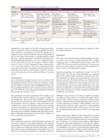
Conventions, with specific medical education and training. the field. We scored the videos on recording clarity, gain, bright-
ness, anatomy, and vessel characteristics on a scoring sheet (Ta-
Microteaching ble 1). The full videos were assessed instead of stills to gain a
A microteaching course was given to combat medics of the better indication of the sonographer’s performance.
Netherlands Armed Forces by two vascular surgeons. The
course consisted of a theoretical part lasting 10 minutes, which Statistical Analysis
included the basic principles and technique of ultrasonogra- All analyses were performed using IBM SPSS Statistics for
phy, supported by a PowerPoint presentation. Afterwards, Mac, version 27 (IBM Corp., Armonk, NY). The observer and
a 5-minute hands-on demonstration was given on how to performer scores distribution and recording quality scores
achieve recordings of the IVC and PV. were generated. Normality was assessed using the Shapiro-
Wilk test. A Mann-Whitney U test was performed to assess
Materials differences in recording times and scores between NSOCMs
Two Lumify (Philips Medical Systems International B.V., and other participants.
™
Best, The Netherlands) handheld ultrasound devices were
used, connected to a Samsung Galaxy Tab A (generation Results
™
10.5, Samsung, Suwon, South-Korea) or a Panasonic FZ-A2
™
tablet (Panasonic, Kadoma, Japan). The IVC was examined us- Performance Assesment
ing the C5-2 abdominal probe, and the PV was assessed using One participant was excluded due to overqualification, as
the L12-4 linear probe. he was a registered nurse. Except for one participant who
96 | JSOM Volume 25, Edition 3 / Fall 2025