
Page 85 - JSOM Summer 2024
P. 85
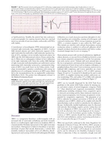
FIGURE 1  (A) The patient’s electrocardiogram (ECG) following a symptomatic period demonstrating sinus bradycardia at a rate of
              59 beats per minute, normal intervals, isolated T-wave inversion in V1 and equivocal T waves in V2 without evidence of epsilon waves.
              (B) An electrocardiogram demonstrating the classic epsilon wave of ARVC in V1. (ECG from ECGpedia.org. distributed under a CC BY-NC-SA
              3.0 DEED license.)  (C) An electrocardiogram demonstrating an epsilon wave in V1 and V2, with T-wave inversions throughout the precordial
                           14
              leads. (ECG from ECGpedia.org. distributed under a CC BY-NC-SA 3.0 DEED license.) 14


              (A)                                (B)                            (C)

              or lightheadedness. Notably, the patient had also undergone   infiltration; as a result, myocytes experience disruption in elec-
              a polysomnography for ongoing insomnia that also reported   trical signaling and contractility, causing arrhythmogenic and
              numerous episodes of narrow-complex tachycardia with PACs   contractile dysfunction.  While various mutations account for
                                                                                   8
              with triplets.                                     ARVC, a common mutation is seen within the  PKP2 gene.
                                                                 This variant can interfere with calcium homeostasis, causing
              A transthoracic echocardiogram (TTE) demonstrated an an-  conduction delays in addition to cell-to-cell adhesion, which
              eurysmal right ventricular base suggestive of ARVC. Cardiac   uniquely produces electrical abnormalities well before cardiac
              MRI showed akinetic  and mildly dyskinetic segment  of the   structural changes may be seen on imaging. 9,10
              right ventricular free wall with a right ventricular ejection frac-
              tion of 52%, meeting a minor morpho-functional criteria for   Most patients present with exertional palpitations, lighthead-
              ARVC per the 2020 International criteria (Padua criteria; Fig-  edness, dyspnea, or syncope from arrhythmias; however, some
              ure 2). There was no radiographic evidence of fatty infiltration   may remain completely asymptomatic until the first presenta-
              of the right ventricular wall. Given his cardiac MRI findings   tion with cardiac arrest.  Patients will rarely present later in
                                                                                    9
              of right ventricular wall abnormalities and ambulatory cardiac   life due to progressive cardiomyopathy and structural changes
              monitoring with ventricular tachycardia, a presumptive diag-  with symptoms of heart failure such as dyspnea, orthopnea, or
              nosis of ARVC was made based on the Padua criteria. Cardi-  peripheral edema. Precordial T-wave inversions, epsilon waves,
              ology recommended a genetics consultation with testing for   widened QRS complexes, and S-wave upstrokes are classic
              identification of a pathogenic variant in an ARVC gene, which   ECG changes but are only found in 30% of ARVC patients
                                                                               8
              led to the recommendation for an implantable cardioverter-   (Figure 1b and 1c).  A normal ECG should not be used to rule
              defibrillator (ICD). The patient was subsequently referred to   out ARVC. A complete workup may include ambulatory car-
              the Medical Evaluation Board.                      diac monitoring, TTE, and cardiac CT or MRI, with referrals
                                                                 to a cardiologist and geneticist, as outlined in Figure 3. 10
              FIGURE 2  Cardiac magnetic resonance imaging with an akinetic and
              mildly dyskinetic segment of the right ventricular free wall along the
              inferior portion of the right ventricular base. The right ventricle was   ARVC  can  be formally  diagnosed  by  the  2010  Task  Force
              normal in size and function with an ejection fraction of 52%.  guidelines or the newer 2020 Padua criteria by meeting ma-
                                                                 jor and minor criteria of several diagnostic categories (Table
                                                                   10
                                                                 1).  Once identified and diagnosed, recommended lifestyle
                                                                 modifications including abstinence from competitive sports
                                                                 or physically provoking activity. Since strenuous exercise pro-
                                                                 motes disease progression, beta-blockers are generally recom-
                                                                                              1
                                                                 mended for sympathetic suppression.  The placement of an
                                                                 ICD is indicated for primary prevention of SCD for all with
                                                                 confirmed cases of ARVC, which can be coupled with class III
                                                                 antiarrhythmics such as sotalol or amiodarone for adjunctive
                                                                 therapy. Additionally, familial implications must be discussed
                                                                 with patients. Interestingly, negative genetic testing does not
                                                                 rule out a diagnosis of ARVC and occurs in 30% of individu-
                                                                                                               11
                                                                 als with an otherwise clear clinical diagnosis of the disease.
                                                                 Therefore, even with inconclusive genetic results, first-degree
              Discussion                                         relatives of ARVC patients should have cardiac surveillance
                                                                 with ECG and ambulatory cardiac monitoring performed ev-
              ARVC  is a  progressive  hereditary  cardiomyopathy  with an   ery 1–3 years after the age of 10 years. 12
              increased risk of ventricular arrhythmia and SCD. Formerly
              known as arrhythmogenic right ventricular dysplasia, ARVC is   While not specifically addressed in the Army Regulation for
              characterized by fibrofatty replacement of the right ventricular   Standards of Medical Fitness (AR 40-501), ARVC presents a
              myocardium.  Pathogenesis is linked to hereditary or sporadic   significant risk of an incapacitating, life-threatening arrhyth-
                        5
              mutations  of  genes  that  encode  desmosomes,  a  protein  that   mia. Subsequently, the Soldier will be placed on a permanent
              normally maintains myocardial cell-to-cell adhesion, detach-  profile and be referred to the Disability Evaluation System
              ing myocytes and causing cell death with subsequent fibrofatty   (DES) to determine if they are retainable, though most will

                                                                                  ARVC in a Special Operations Soldier  |  83