
Page 50 - JSOM Winter 2023
P. 50
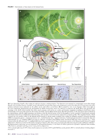
FIGURE 1  Mechanisms of blast injury in the human brain.


                                                                                                   Artwork in Panels A and B by Kimberly Main Knoper.


                          Inflammation      Astroglial Scarring    Axonal Injury   Tau Deposition


                       IBA1            GFAP                     ß- APP          AT8

          (A) Four Operators breach a door using an explosive during a combat mission. The Operators are exposed to overpressure waves that emerge
          directly from the blast location (incident waves indicated by curved white lines) and overpressure waves that rebound off the ground and other
          surfaces (reflected waves indicated by the yellow arrow and yellow curved lines). (B) A zoomed-in view of an Operator (third from right in
          the stack formation) demonstrates the mechanisms by which overpressure waves are believed to penetrate the skull. Waves may penetrate the
          openings in the skull—which include the orbits, nasal passages, oral cavity, ear canals, and foramen magnum—resulting in focal brain injury.
          Overpressure waves may also penetrate the helmet and skull, causing diffuse brain injury. The left hemisphere of the Operator’s brain is shown in
          the sagittal plane, with the right hemisphere not shown so that the medial surface of the left hemisphere can be visualized. Default mode network
          functional connectivity is displayed in yellow/orange, and executive control network connectivity is shown in blue. The cingulum bundle, which
          connects the core midline nodes of the default mode network, is shown using standard color-coding for diffusion magnetic resonance imaging
          (MRI) tractography (green = anterior-posterior; red = medial-lateral; blue = superior-inferior). Functional and structural connectivity data are
          superimposed on a T1-weighted MRI scan from an Operator enrolled in the ReBlast study. Functional MRI data were acquired on a 7 Tesla
          scanner, while the diffusion and T1-weighted MRI data were acquired on the 3 Tesla Connectome scanner at the Massachusetts General Hospital
                                                                                                            71
          Athinoula A. Martinos Center for Biomedical Imaging in Boston, MA. Data were processed and analyzed using previously published methods.
          (C) Representative pathology (arrows) detected by immunohistochemistry in the brains of military personnel exposed to repeated blasts includes
          inflammation, astroglial scarring, axonal injury, and tau deposition.
          AT8 = anti-phospho-tau; ß-APP = beta-amyloid precursor protein; GFAP = glial fibrillary acidic protein; IBA1 = ionized calcium binding adaptor
          molecule 1.

          48  |  JSOM   Volume 23, Edition 4 / Winter 2023