
Page 104 - JSOM Summer 2022
P. 104
patients with moderate to severe ARDS utilizing the high PEEP Adjunctive Equipment
20
ARDSnet strategy. Adjuncts including tube thoracostomy
may be required with mechanical ventilation. Overall, most A bag-valve mask (BVM), suction catheter (Yankauer or
thoracic injury trauma cases are managed nonoperatively. DuCanto), oral and nasal airways, and oxygen source are the
minimum stockage for a provider with a mechanical ventila-
tion capability. The ventilator may allow the medic or pro-
Individualization of Ventilator Settings vider to perform other functions in a patient with respiratory
failure ordinarily requiring BVM treatment. Prehospital or
Establishing initial settings for the mechanical ventilator is
straightforward in most patients requiring ventilation. The transport ventilators used by SOF medics and providers are
plurality of patients with profound hypoxic or ventilatory de- traditionally smaller and less feature available than their hos-
fects can be effectively managed by an algorithmic approach pital-based counterparts. Sophisticated turbine-based designs
to mechanical ventilation, adjusting to the clinical situation. have now largely replaced pneumatic ventilator units. Most of
1. Establish Ventilator Mode these ventilators have an established ability to maintain ade-
o Start with Volume Assist-Control. quate SpO in severe acute hypoxic respiratory failure animal
2
2. Set Tidal Volume (TV) models and should be regarded as sufficient to treat even the
24
o Calculate the ideal body weight (IBW) in kilograms. ARDS patient.
This is not the patient’s actual body weight, despite hav-
ing a fit physique. Continuous Monitoring Techniques
– IBW (male) (kg) = 50 + 2.3 (height in inches – 60)
– IBW (female) (kg) = 45.5 + 2.3 (height in inches – 60) Approximately 97% of the capacity of delivered oxygen is
o Select a tidal volume of 7cc/kg multiplied by the IBW. bound to the hemoglobin unit. Therefore, pulse oximetry
o Ultimately, target a 6–8cc/kg IBW goal. of the peripheral saturation of O (SpO ) is the mainstay of
2
2
3. Set the Respiratory Rate (RR) monitoring for patients receiving mechanical ventilation.
o Start with a respiratory rate of 18 breaths per minute. More accurate and direct measurement of SpO PaO , and
2
2,
Normal ventilation rates run between 10 and 22. PaCO can be accomplished by point-of-care oximetry in a
2
o Change RR instead of TV to change patient’s minute prehospital situation but are not essential. Capnography and/
ventilation. or end-tidal CO (EtCO ) can provide capability for monitor-
2
2
o Adjust RR to achieve pH > 7.25 to 7.30 or PaCO 35–45 ing ventilation factors and are considered the gold standard of
2 monitoring tube placement and ventilation status. Indepen-
25
or ETCO 35–45. As tidal volume is restricted in prac-
2
ticing LTVV, you may require advancing the RR into the dent variables of ventilation should be monitored frequently:
30s in order to achieve respiratory compensation. peak and plateau airway pressures should be monitored in
4. Oxygenate the Patient volume-controlled modes, while TV should be observed when
o Start FiO of 100%. pressure is being controlled. Sedation should be examined by
2 validated tools such as the Richmond Agitation-Sedation Scale
o Start PEEP at 5 for patients without significant hypoxia
prior to intubation. (RASS) as undersedation may contribute to patient–ventilator
o Alternatively, start PEEP at 10 for hypoxic patients. asynchrony
o Decrease FiO by 10% every 2–5 minutes as to keep
2
SpO > 90%. Troubleshooting
2
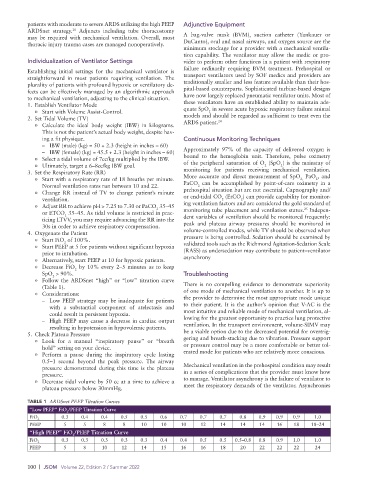
o Follow the ARDSnet “high” or “low” titration curve
(Table 1). There is no compelling evidence to demonstrate superiority
o Considerations: of one mode of mechanical ventilation to another. It is up to
– Low PEEP strategy may be inadequate for patients the provider to determine the most appropriate mode unique
with a substantial component of atelectasis and to their patient. It is the author’s opinion that V-AC is the
could result in persistent hypoxia. most intuitive and reliable mode of mechanical ventilation, al-
– High PEEP may cause a decrease in cardiac output lowing for the greatest opportunity to practice lung protective
resulting in hypotension in hypovolemic patients. ventilation. In the transport environment, volume-SIMV may
5. Check Plateau Pressure be a viable option due to the decreased potential for overtrig-
o Look for a manual “inspiratory pause” or “breath gering and breath-stacking due to vibration. Pressure support
hold” setting on your device. or pressure control may be a more comfortable or better tol-
o Perform a pause during the inspiratory cycle lasting erated mode for patients who are relatively more conscious.
0.5–1 second beyond the peak pressure. The airway
pressure demonstrated during this time is the plateau Mechanical ventilation in the prehospital condition may result
pressure. in a series of complications that the provider must know how
o Decrease tidal volume by 50 cc at a time to achieve a to manage. Ventilator asynchrony is the failure of ventilator to
plateau pressure below 30mmHg. meet the respiratory demands of the ventilator. Asynchronies
TABLE 1 ARDSnet PEEP Titration Curves
“Low PEEP” FiO /PEEP Titration Curve
2
0.3 0.4 0.4 0.5 0.5 0.6 0.7 0.7 0.7 0.8 0.9 0.9 0.9 1.0
FiO 2
PEEP 5 5 8 8 10 10 10 12 14 14 14 16 18 18–24
“High PEEP” FiO /PEEP Titration Curve
2
0.3 0.3 0.3 0.3 0.3 0.4 0.4 0.5 0.5 0.5–0.8 0.8 0.9 1.0 1.0
FiO 2
PEEP 5 8 10 12 14 15 16 16 18 20 22 22 22 24
100 | JSOM Volume 22, Edition 2 / Summer 2022