
Page 102 - JSOM Summer 2022
P. 102
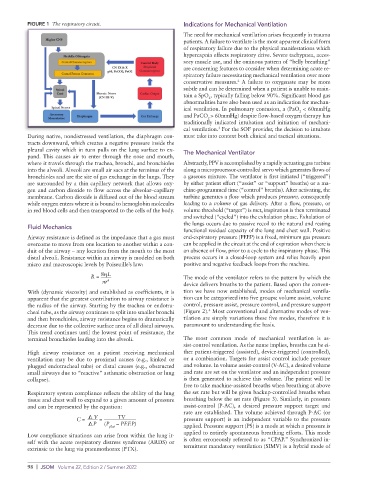
FIGURE 1  The respiratory circuit.                 Indications for Mechanical Ventilation
                                                             The need for mechanical ventilation arises frequently in trauma
                                                             patients. A failure to ventilate is the most apparent clinical form
                                                             of respiratory failure due to the physical manifestations which
                                                             hypercapnia effects respiratory drive. Severe tachypnea, acces-
                                                             sory muscle use, and the ominous pattern of “belly breathing”
                                                             are concerning features to consider when determining acute re-
                                                             spiratory failure necessitating mechanical ventilation over more
                                                             conservative measures.  A failure to oxygenate may be more
                                                                               2
                                                             subtle and can be determined when a patient is unable to main-
                                                             tain a SpO , typically falling below 90%. Significant blood gas
                                                                     2
                                                             abnormalities have also been used as an indication for mechan-
                                                             ical ventilation. In pulmonary contusion, a (PaO  < 60mmHg
                                                                                                   2
                                                             and PaCO > 60mmHg) despite flow-based oxygen therapy has
                                                                     2
                                                             traditionally indicated intubation and initiation of mechani-
                                                             cal ventilation.  For the SOF provider, the decision to intubate
                                                                        3
          During native, nondistressed ventilation, the diaphragm con-  must take into context both clinical and tactical situations.
          tracts downward, which creates a negative pressure inside the
          pleural cavity which in turn pulls on the lung surface to ex-
          pand. This causes air to enter through the nose and mouth,   The Mechanical Ventilator
          where it travels through the trachea, bronchi, and bronchioles   Abstractly, PPV is accomplished by a rapidly actuating gas turbine
          into the alveoli. Alveoli are small air sacs at the terminus of the   along a microprocessor-controlled servo which generates flows of
          bronchioles and are the site of gas exchange in the lungs. They   a gaseous mixture. The ventilator is first initiated (“triggered”)
          are surrounded by a thin capillary network that allows oxy-  by either patient effort (“assist” or “support” breaths) or a ma-
          gen and carbon dioxide to flow across the alveolar–capillary   chine-programmed time (“control” breaths). After activating, the
          membrane. Carbon dioxide is diffused out of the blood stream   turbine generates a flow which produces pressure, consequently
          while oxygen enters where it is bound to hemoglobin molecules   leading to a volume of gas delivery. After a flow, pressure, or
          in red blood cells and then transported to the cells of the body.  volume threshold (“target”) is met, inspiration is then terminated
                                                             and switched (“cycled”) into the exhalation phase. Exhalation of
                                                             the lungs occurs due to passive recoil to the natural and resting
          Fluid Mechanics
                                                             functional residual capacity of the lung and chest wall. Positive
          Airway resistance is defined as the impedance that a gas must   end-expiratory pressure (PEEP) is a fixed, minimum gas pressure
          overcome to move from one location to another within a con-  can be applied in the circuit at the end of expiration when there is
          duit of the airway – any location from the mouth to the most   an absence of flow, prior to a cycle to the inspiratory phase. This
          distal alveoli. Resistance within an airway is modeled on both   process occurs in a closed-loop system and relies heavily upon
          micro and macroscopic levels by Poiseuille’s law:  positive and negative feedback loops from the machine.

                               R =  8ηL                      The mode of the ventilator refers to the pattern by which the
                                  πr 4
                                                             device delivers breaths to the patient. Based upon the conven-
          With (dynamic viscosity) and established as coefficients, it is   tion we have now established, modes of mechanical ventila-
          apparent that the greatest contribution to airway resistance is   tion can be categorized into five groups: volume assist, volume
          the radius of the airway. Starting by the trachea or endotra-  control, pressure assist, pressure control, and pressure support
                                                                     4
          cheal tube, as the airway continues to split into smaller bronchi   (Figure 2).  Most conventional and alternative modes of ven-
          and then bronchioles, airway resistance begins to dramatically   tilation are simply variations these five modes, therefore it is
          decrease due to the collective surface area of all distal airways.   paramount to understanding the basis.
          This trend continues until the lowest point of resistance, the
          terminal bronchioles leading into the alveoli.     The most common mode of mechanical ventilation is as-
                                                             sist-control ventilation. As the name implies, breaths can be ei-
          High airway resistance on a patient receiving mechanical   ther patient-triggered (assisted), device-triggered (controlled),
          ventilation  may  be due  to  proximal  causes  (e.g.,  kinked or   or a combination. Targets for assist control include pressure
          plugged endotracheal tube) or distal causes (e.g., obstructed   and volume. In volume assist-control (V-AC), a desired volume
          small airways due to “reactive” asthmatic obstruction or lung   and rate are set on the ventilator and an independent pressure
          collapse).                                         is then generated to achieve this volume. The patient will be
                                                             free to take machine-assisted breaths when breathing at above
          Respiratory system compliance reflects the ability of the lung   the set rate but will be given backup-controlled breaths when
          tissue and chest wall to expand to a given amount of pressure   breathing below the set rate (Figure 3). Similarly, in pressure
          and can be represented by the equation:            assist-control (P-AC), a desired pressure support target and
                                                             rate are established. The volume achieved through P-AC (or
                                ∇  V  =           TV         pressure support) is an independent variable to the pressure
                                      P     (P  – PEEP)
                                     plat                    applied. Pressure support (PS) is a mode at which a pressure is
                          C =  ∇
                                                             applied to entirely spontaneous breathing efforts. This mode
          Low compliance situations can arise from within the lung it-
          self with the acute respiratory distress syndrome (ARDS) or   is often erroneously referred to as “CPAP.” Synchronized in-
          extrinsic to the lung via pneumothorax (PTX).      termittent mandatory ventilation (SIMV) is a hybrid mode of

          98  |  JSOM   Volume 22, Edition 2 / Summer 2022