
Page 115 - 2022 Spring JSOM
P. 115
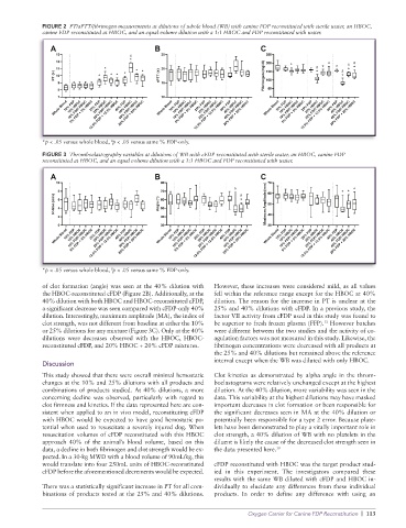
FIGURE 2  PT/aPTT/fibrinogen measurements at dilutions of whole blood (WB) with canine FDP reconstituted with sterile water, an HBOC,
              canine FDP reconstituted at HBOC, and an equal volume dilution with a 1:1 HBOC and FDP reconstituted with water.


              *p < .05 versus whole blood,  p < .05 versus same % FDP-only.
                                  δ
              FIGURE 3  Thromboelastography variables at dilutions of WB with cFDP reconstituted with sterile water, an HBOC, canine FDP
              reconstituted at HBOC, and an equal volume dilution with a 1:1 HBOC and FDP reconstituted with water.


              *p < .05 versus whole blood,  p < .05 versus same % FDP-only.
                                  δ
              of clot formation (angle) was seen at the 40% dilution with   However, these increases were considered mild, as all values
              the HBOC-reconstituted cFDP (Figure 2B). Additionally, at the   fell within the reference range except for the HBOC at 40%
              40% dilution with both HBOC and HBOC-reconstituted cFDP,   dilution. The reason for the increase in PT is unclear at the
              a significant decrease was seen compared with cFDP-only 40%   25% and 40% dilutions with cFDP. In a previous study, the
              dilution. Interestingly, maximum amplitude (MA), the index of   factor VII activity from cFDP used in this study was found to
                                                                                                 13
              clot strength, was not different from baseline at either the 10%   be superior to fresh frozen plasma (FFP).  However batches
              or 25% dilutions for any mixture (Figure 3C). Only at the 40%   were different between the two studies and the activity of co-
              dilutions were decreases observed with the HBOC, HBOC-   agulation factors was not measured in this study. Likewise, the
              reconstituted cFDP, and 20% HBOC + 20% cFDP mixtures.  fibrinogen concentrations were decreased with all products at
                                                                 the 25% and 40% dilutions but remained above the reference
                                                                 interval except when the WB was diluted with only HBOC.
              Discussion
              This study showed that there were overall minimal hemostatic   Clot kinetics as demonstrated by alpha angle in the throm-
              changes at the 10% and 25% dilutions with all products and   boelastograms were relatively unchanged except at the highest
              combinations of products studied. At 40% dilutions, a more   dilution. At the 40% dilution, more variability was seen in the
              concerning decline was observed, particularly with regard to   data. This variability at the highest dilutions may have masked
              clot firmness and kinetics. If the data represented here are con-  important decreases in clot formation or been responsible for
              sistent when applied to an in vivo model, reconstituting cFDP   the significant decreases seen in MA at the 40% dilution or
              with HBOC would be expected to have good hemostatic po-  potentially been responsible for a type 2 error. Because plate-
              tential when used to resuscitate a severely injured dog. When   lets have been demonstrated to play a vitally important role in
              resuscitation volumes of cFDP reconstituted with this HBOC   clot strength, a 40% dilution of WB with no platelets in the
              approach 40% of the animal’s blood volume, based on this   diluent is likely the cause of the decreased clot strength seen in
              data, a decline in both fibrinogen and clot strength would be ex-  the data presented here. 19
              pected. In a 30-kg MWD with a blood volume of 90mL/kg, this
              would translate into four 250mL units of HBOC-reconstituted   cFDP reconstituted with HBOC was the target product stud-
              cFDP before the aforementioned decrements would be expected.  ied in this experiment. The investigators compared these
                                                                 results with the same WB diluted with cFDP and HBOC in-
              There was a statistically significant increase in PT for all com-  dividually to elucidate any differences from these individual
              binations of products tested at the 25% and 40% dilutions.   products. In order to define any difference with using an

                                                                           Oxygen Carrier for Canine FDP Reconstitution  |  113