
Page 82 - 2020 JSOM Winter
P. 82
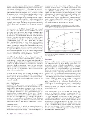
minutes  after  the  induction  of  CA.  As  soon  as  ECMO  was   increased from 0 (–2 to +2) to 29 (28 to 30), and the pH level
          initiated, we stopped the external compressions. In animal No.   gradually decreased from 7.25 (7.18 to 7.48) to 6.66 (6.50
          1, both short cannulae (a 12-Fr 9" arterial and an 18-Fr 12"   to 6.82) during the time course (Figure 5). Despite resusci-
          venous) were inserted into the left femoral vessels using an   tation, ECMO, and blood replacement, rapidly developing
          open cutdown exposure. In animal No. 2, ultrasound-guided   hyper kalemia was observed in the two study animals. In these
          percutaneous access was initially attempted to achieve venous   study subjects, the potassium level increased from 2.8mmol/L
          and arterial sheath placement (a 12-Fr 7.5" arterial and a 17-  (2.0–3.8mmol/L) to 7.8mmol/L (6.8–8.8mmol/L). In addition,
          Fr 30"). Body movements during the CPR and small-caliber   these two study animals demonstrated  a dramatic  and pro-
          vessels hindered the ability to achieve a stable needle position,   gressive decrease in hemoglobin levels to the end of the study
          however, so access was rapidly transitioned to a semi-Seldinger   (Figure 5). No blood samples were taken from animal No. 3
          technique that involved cutdown to expose the anterior vessel   after aortic cannulation and initiation of ECMO.
          walls for direct needle puncture of both the femoral artery and
          vein.                                              FIGURE 5  Blood gases analysis summarizing three study animals
                                                             that underwent extracorporeal cardiopulmonary resuscitation. Data
                                                             presented as mean (standard error of mean).
          After connection to the ECMO circuit, E-CPR was initiated
          with a BFR of 2.0–2.5L/min, and the ventilator was discon-
          nected. The two study animals were initially resuscitated with
          9L and 4L of crystalloids, respectively. This was followed by
          the infusion of 1L of stored whole blood and 10mg of calcium
          chloride. Thereafter, the two animals were transported with
          ongoing ECMO by helicopter to the Role 2 facility. After a
          15-minute flight to the Role 2 and 3 hours after CA, animal
          No. 1 developed ACS due to severe blood loss (hemoglobin
          level decreased from 10.2 to 1.3g/dL), shock (MAP decreased
          from 97 to 34mmHg), and extensive fluid replacement. The re-
          sulting ACS contributed to inferior vena cava compression and
          inadequate blood drainage via the short-access venous cannula,
          which resulted in a drop of the ECMO circuit BFR to 400mL/
          min. An emergent decompressive laparotomy was subsequently
          peformed at the Role 2, restoring the BFR to 1.5L/min.
                                                                                                   +
                                                             BD = base deficit; Hb = hemoglobin (g/dL) level; K  = potassium
                                                             (mmol/L), pH level.
          Animal No. 2 received a lower volume of crystalloids and de-
          veloped no complications en route to the Role 2, although a   Discussion
          similar drop in measured hemoglobin level was observed (9.5
          to 1.5g/dL). Despite effective blood drainage and return, this   Extensive combat trauma is a leading cause of prehospital
          second study animal continued to deteriorate and developed   and  in-hospital mortality  in the  warfare  environment.  Ex-
          progressive shock (MAP decrease from 65 to 22mmHg). Ad-  sanguination leading to blood volume depletion remains an
          ditional carotid artery cannulation was attempted to restore   ever-present potential challenge. Despite advances in TCCC,
          BFR at the Role 2 via the addition of two arterial return can-  ARC, far- forward damage-control surgery, and resuscitation,
          nulae, with no notable effect.                     there remains a need to evaluate the utility of further emerging
                                                             techniques capable of supporting and restoring circulation in
          Perfusion of both animals was artifically maintained during   casualties with TCA. E-CPR (V-A ECMO during CA), already
          the experimental protocol in its entirety. The protocol was ter-  shown to be an effective tool in the rescue from cardiogenic
          minated in both study animals after 4 hours without ROSC.   CA in select patients, 10,14  warrants examination in this regard.
          We recorded no access-related complications in either of the   However, E-CPR for acute trauma remains a controversial
          two study animals.                                 topic requiring additional study. Although ongoing hemor-
                                                             rhage is considered to be a traditional contraindication for
          Role 1 E-CPR Failure                               ECMO due to the typically required systemic heparinization,
          In animal No. 3, open femoral vein cannulation (an 18-Fr 12"   some civilian trauma centers have already demonstrated the
          cannula)  was  successfully  achieved  17  minutes  after  the  in-  potential for saving lives using this technique, even for poly-
          duction of CA. Arterial access via cutdown of the femoral, ca-  traumatized patients. 14
          rotid, and even iliac arteries (a 12-Fr 9" cannula) was not able
          to be achieved, however, because of profound spasm and hy-  Early hospital-based use of V-A ECMO has already been
          potension. Open aortic cannulation via laparotomy ultimately   found to be effective for refractory nontraumatic CA.  In
                                                                                                         15
          enabled the circuit to commence at a BFR of 1L/min. The arte-  order to explore the potential value of this adjunct as early
          rial cannulae at this location were not able to be appropriately   after arrest as possible, the use of V-A ECMO has also been
                                                                                                       16
          secured for transport, however. Because of these challenges   pushed forward for potential prehospital applications.  These
          and extensive bleeding from the aortic cannulation sites, the   investigations have shown that the interval between CA and
          protocol for the third animal was discontinued due to futility.  restoration of circulation (the low-flow period) is inversely as-
                                                             sociated with optimal neurologic and clinical outcome after
                                                                   17
          Laboratory Values                                  E-CPR.  To optimize out-of-hospital care and reduce the low-
          Blood tests taken from all animals demonstrated a dramatic   flow time period, special ECMO teams have been developed
          progression of metabolic acidosis because of blood loss and   in some countries. 16,18  ECMO experience in austere military
          extensive fluid replacement therapy. The base deficit gradually   circumstances is, however, limited.


          80  |  JSOM   Volume 20, Edition 4 / Winter 2020