
Page 80 - 2020 JSOM Winter
P. 80
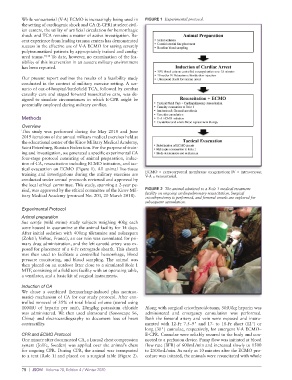
While venoarterial (V-A) ECMO is increasingly being used in   FIGURE 1  Experimental protocol.
          the setting of cardiogenic shock and CA (E-CPR) at select civil-
          ian centers, the utility of artificial circulation for hemorrhagic
          shock and TCA remains a matter of active investigation. Re-
          cent experience from leading trauma centers has demonstrated
          success in the effective use of V-A ECMO for saving severely
          polytraumatized patients by appropriately trained and config-
          ured teams. 10,14  To date, however, no examination of the fea-
          sibility of this intervention in an austere military environment
          has been reported.
          Our present report outlines the results of a feasibility study
          conducted in the context of military exercise setting. A sce-
          nario of out-of-hospital/battlefield TCA, followed by combat
          casualty care and staged forward resuscitative care, was de-
          signed to simulate circumstances in which E-CPR might be
          potentially employed during military conflict.


          Methods
          Overview
          This study was performed during the May 2018 and June
          2019 iterations of the annual military medical exercises held at
          the educational center of the Kirov Military Medical Academy,
          Saint Petersburg, Russian Federation. For the purpose of train-
          ing and investigation, we generated a specific experimental CA
          four-stage protocol consisting of animal preparation, induc-
          tion of CA, resuscitation including ECMO initiation, and tac-
          tical evacuation on ECMO (Figure 1). All animal live-tissue
          training and investigations during the military exercises are   ECMO = extracorporeal membrane oxygenation; IV = intravenous;
                                                             V-A = venoarterial.
          conducted under annual protocols reviewed and approved by
          the local ethical committee. This study, spanning a 2-year pe-
          riod, was approved by the ethical committee of the Kirov Mil-  FIGURE 2  The animal admitted to a Role 1 medical treatment
          itary Medical Academy (protocol No. 203, 20 March 2018).  facility on ongoing cardiopulmonary resuscitation. Surgical
                                                             cricothyrotomy is performed, and femoral vessels are explored for
                                                             subsequent cannulation.
          Experimental Protocol
          Animal preparation
          Sus scrofa (wild swine) study subjects weighing 40kg each
          were housed in quarantine at the animal facility for 14 days.
          After initial sedation with 400mg tiletamine and zolazepam
          (Zoletil; Virbac, France), an ear vein was cannulated for pri-
          mary drug administration, and the left carotid artery was ex-
          posed for placement of a 6-Fr retrograde sheath. This sheath
          was then used to facilitate a controlled hemorrhage, blood
          pressure  monitoring,  and  blood sampling.  The  animal was
          then placed on an outdoor litter close to a simulated Role 1
          MTF, consisting of a field tent facility with an operating table,
          a ventilator, and a basic kit of surgical instruments.

          Induction of CA
          We chose a combined (hemorrhage-induced plus nontrau-
          matic) mechanism of CA for our study protocol. After con-
          trolled removal of 35% of total blood volume (stored using
          1000IU of heparin per unit), 20mg/kg potassium chloride   Along with surgical cricothyroidotomy, 50IU/kg heparin was
          was  administered.  We then  used  ultrasound  (Sonoscape  S6,   administered and emergency cannulation was performed.
          China)  and electrocardiography  to document  loss  of  heart   Both the femoral artery and vein were exposed and instru-
          contractility.                                     mented  with  12-Fr  7.5–9"  and  17-  to  18-Fr  short  (12")  or
                                                             long (30") cannulae, respectively, for emergent V-A ECMO–
          CPR and ECMO Protocol                              E-CPR. Cannulae were reliably secured to the body and con-
          One minute after documented CA, a Lucas2 chest compression   nected to a perfusion device. Pump flow was initiated at blood
          system (Jolife, Sweden) was applied over the animal’s chest   flow rate (BFR) of 600mL/min and increased slowly to 1500
          for ongoing CPR. During CPR, the animal was transported   to 2500mL/min. As early as 10 minutes after the ECMO pro-
          to a tent (Role 1) and placed on a surgical table (Figure 2).    cedure was initated, the animals were resuscitated with whole


          78  |  JSOM   Volume 20, Edition 4 / Winter 2020