
Page 81 - 2020 JSOM Winter
P. 81
blood. To avoid severe hypocalcemia, small boluses of 10% Study Termination and Euthanasia
calcium chloride were administered. Epinephrine was used to After delivery to the Role 2, animals were checked for MAP
correct critical hypotension. and possible return of spontaneous circulation (ROSC). If
indicated, the swine underwent additional appropriate inter-
Blood samples were taken for gas analisys (i-Stat, Abbott Lab- vention for stabilization and continuous monitoring, such as
oratories, IL) at the following timepoints: baseline, 5 minutes abdominal compartment syndrome (ACS) release or/and ad-
after CA (pre-ECMO), 1 hour after ECMO initiation (on- ditional vascular cannulation. If no ROSC was achieved after
ECMO), and on admission to Role 2 before study termination these procedures, death was confirmed, the study was termi-
(terminal-ECMO). Mean arterial pressure (MAP), flow, and nated, and the ECMO circuit stopped. No postmortem exam-
saturation parameters were monitored throughout the study. ination was undertaken.
The primary endpoint was the adequate level of perfusion
pressure on admission to Role 2.
Results
One experienced anesthetist-perfusionist responsible for Overview
ECMO augmented a Role 1 team consisting of an anesthe- Three sedated animals underwent induction of CA at a dis-
tist, a military surgeon, and a scrub nurse. The air critical care tance of 100–150 m from the Role 1 facility and developed
transport team consisted of another anesthetist, the anesthe- sustained asystole. On the scene, care providers initated intra-
tist-perfusionist joining the transport, and two anesthetist osseus fluid replacement (200mL Ringer solution) and CPR
nurses equipped with a dedicated trauma care package. The within 1.4 minutes (range, 1–2 minutes), followed by immedi-
military surgeon experienced in performing REBOA and other ate ground transportation to the Role 1, which took 5 minutes.
basic endovascular interventions in human and animals, but Because of low oxygen saturations (<60%) upon admission,
having no experience in large cannulae insertion, performed all surgical cricothyroidotomy was performed with high-flow
cannulations in our study. No additional training for ECMO 100% oxygen administered via a tracheostomy tube. Lucas
in animals was undertaken before the study began. chest compressions supported effective but gradually deterio-
rating blood circulation, confirmed by decreasing MAP from
Field ECMO Equipment initial values of 55mmHg (range, 50–65mmHg) to 40mmHg
For the ECMO circuit, we used a lightweight (3kg) portable (range, 20–40mmHg) prior to ECMO initiation (Figure 4).
perfusion system, Ex-Stream (TransBiotech, Ltd., Skolkovo, Two of three animals underwent immediate successful can-
Russian Federation). The whole ECMO kit, weighing approx- nulation and ECMO initiation, resulting in restoration of
imately 3–4kg (total kit dimensions, 50 × 40 × 25cm), also in- perfusion pressure to 80mmHg (range, 70–90mmHg). We ulti-
cludes an oxygenator (Affinity Pixie, Medtronic, Fridley, MN), mately evacuated these two animals to the Role 2, followed by
venous (access) and arterial (return) cannulae, a spare ECMO additional surgical interventions and study termination. The
circuit, connectors, tubes, sterile scissors, and tubing clamps. arteries of animal No. 3 were unable to be cannulated after
The ECMO circuit was primed and prepared. Bio-Medicus multiple attempts over an hour because of spastic small-caliber
and DLP pediatric cannulae were used for semi-Seldinger and vessels. In this latter animal, the protocol was ultimately dis-
open cannulation, respectively (all Medtronic). continued due to futility. No ROSC was ultimately achieved in
either of the remaining study animals.
Evacuation Protocol
Once the ECMO circuit was stabilized, the animal underwent Successful Role 1 E-CPR
immediate evacuation by a rotary wing platform to a Role 2 Two animals underwent a complete protocol of ECMO initia-
MTF deployed in fast adjustable pneumatic modules within tion under ongoing CPR. Cannulation of femoral vessels was
the distance of a 15-minute flight (Figure 3). A single, dedi- performed within 20 and 18 minutes after arrival to Role 1,
cated, high-capacity Mil Mi-8 helicopter equipped with a stan- respectively, and restoration of flow was achieved 25 and 23
dard medical transportation module was used for the flights.
FIGURE 4 Diagram illustrating perfusion pressure over the study
time course. Data presented as mean (standard error of mean).
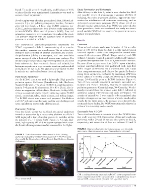
FIGURE 3 Tactical evacuation of the animal to Role 2 during
extracorporeal cardiopulmanory resuscitation (V-A ECMO) using
the portable Ex-Stream device (TransBiotech, Ltd, Skolkovo, Russian
Federation).
CPR = cardiopulmonary resuscitation; ECMO = extracorporeal mem-
brane oxygenation; E-CPR = extracorporeal cardiopulmonary resusci-
V-A = venoarterial. tation; MAP = mean arterial pressure; POI = point of injury.
Battlefield Extracorporeal Cardiopulmonary Resuscitation | 79