
Page 38 - 2020 JSOM Winter
P. 38
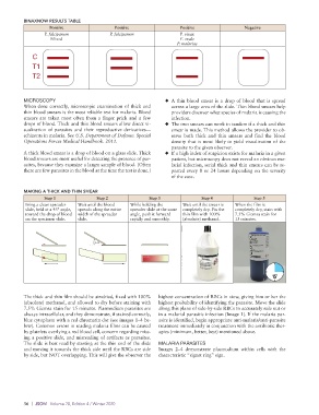
BINAXNOW RESULTS TABLE
                    Positive                Positive                 Positive                Negative
                  P. falciparum            P. falciparum             P. vivax
                    Mixed                                            P. ovale
                                                                    P. malariae
             C

             T1
             T2


          MICROSCOPY                                           ◆   A thin blood smear is a drop of blood that is spread
          When done correctly, microscopic examination of thick and   across a large area of the slide. Thin blood smears help
          thin blood smears is the most reliable test for malaria. Blood   providers discover what species of malaria is causing the
          smears are taken most often from a finger prick and a few   infection.
          drops of blood. Thick and thin blood smears allow direct vi-  ◆   The two smears can work in tandem if a thick and thin
          sualization of parasites and their reproductive derivatives—  smear is made. This method allows the provider to ob-
          schizonts in malaria. See U.S. Department of Defense. Special   serve both thick and thin smears and find the blood
          Operations Forces Medical Handbook. 2011.               density that is most likely to yield visualization of the
                                                                  parasite to the given observer.
          A thick blood smear is a drop of blood on a glass slide. Thick   ◆   If a high index of suspicion exists for malaria in a given
          blood smears are most useful for detecting the presence of par-  patient, but microscopy does not reveal an obvious ma-
          asites, because they examine a larger sample of blood. (Often   larial infection, serial thick and thin smears can be re-
          there are few parasites in the blood at the time the test is done.)  peated every 8 or 24 hours depending on the severity
                                                                  of the case.

          MAKING A THICK AND THIN SMEAR
                  Step 1              Step 2             Step 3              Step 4              Step 5
           Bring a clean spreader   Wait until the blood   While holding the   Wait until the smear is   When the film is
           slide, held at a 45° angle,   spreads along the entire   spreader slide at the same   completely dry. Fix the   completely dry, stain with
           toward the drop of blood   width of the spreader   angle, push it forward   thin film with 100%   7.5% Giemsa stain for
           on the specimen slide.  slide.         rapidly and smoothly.  (absolute) methanol.  15 minutes.


          The thick and thin film should be air-dried, fixed with 100%   highest concentration of RBCs in view, giving him or her the
          (absolute) methanol, and allowed to dry before staining with   highest probability of identifying the parasite. Move the slide
          7.5% Giemsa stain for 15 minutes. Plasmodium parasites are   along this plane of side-by-side RBCs to accurately rule out or
          always intracellular, and they demonstrate, if stained correctly,   in a malarial parasitic infection (Image 1). If the malaria par-
          blue cytoplasm with a red chromatin dot (see images 1–4 be-  asite is identified, begin appropriate anti-malaria/anti- parasite
          low). Common errors in reading malaria films can be caused   treatment immediately in conjunction with the antibiotic ther-
          by platelets overlying a red blood cell, concern regarding miss-  apies (minimum, better, best) mentioned above.
          ing a positive slide, and misreading of artifacts as parasites.
          The slide is best read by starting at the thin end of the slide   MALARIA PARASITES
          and moving it towards the thick side until the RBCs are side   Images 2–4 demonstrate plasmodium within cells with the
          by side, but NOT overlapping. This will give the observer the   characteristic “signet ring” sign.


          36  |  JSOM   Volume 20, Edition 4 / Winter 2020