
Page 132 - 2020 JSOM Winter
P. 132
returned to normal 1 hour postexposure. In deep frostbite, may improve. Areas that do not take up the tracer in delayed
skin temperature determined by thermography was near am- images will likely require amputation (specificity = 0.99, sen-
bient temperature at 1 hour postexposure and for at least 1 sitivity = 0.92). 83,84
week postexposure (i.e., end of study). Further research on
78
humans is needed, but thermography may be a promising The addition of SPECT to multiphase 99m Tc bone scintigraphy
technique to determine the severity of frostbite injury. can improve diagnosis by identifying the precise anatomic level
of bone necrosis. SPECT images are obtained immediately
Other imaging techniques are more appropriate in hospital after acquiring bone scan delayed-phase images. The three-
environments. Digital subtraction angiography (DSA) is a flu- dimensional CT scans are viewed either side-by-side with the
oroscopic technique that uses radiography to obtain real-time bone scans or combined by some commercial imagers. This
moving images of the blood vessels while masking out other provides precise anatomic identification of the demarcation
structures (e.g., bone). It requires injection of a contrast dye. It line between viable and nonviable bone, allowing for more
is useful for patients presenting within 24 hours of a deep frost- accurate surgical planning. 68,79,85
bite injury with suspected blood vessel involvement because it
assesses blood vessel patency. 68,79 It has been suggested that Long-Term Sequelae
68
DSA might also be used to identify targets for thrombolytic The most common long-term sequela from frostbite is a gen-
agents and to determine the progress and effectiveness of this eral hypersensitivity to cold in the affected area or areas. 86–89
treatment. Other persistent symptoms among frostbitten patients include
hyperhidrosis, decreased tactile sensitivity, and pain. 86–89 Com-
Magnetic resonance imaging angiography is a noninvasive al- pared with unaffected areas, formerly frostbitten areas reach
ternative to DSA that is also useful for examining blood vessel a lower skin temperature on exposure to cold. In frostbitten
88
patency and assisting in determining the level of tissue loss. toes and fingers, CIVD is delayed or abolished 3 to 4 years
It allows precise visualization of blood vessels and provides a after the injury, suggesting higher susceptibility to future cold
demarcation line between viable and nonviable tissue that can injury. Among 40 soldiers followed up 6 months after a frost-
90
assist in surgical planning. 80,81 Some clinicians state that the bite injury, 26 (65%) had symptoms consistent with neurovas-
utility of this technique has limitations. 68 cular injury, including cold sensitivity paresthesia, pain, and
hyperesthesia, as well as lower tolerance for cold water expo-
Bone scintigraphy involves injection of a low-level radioactive sure. Ninety-seven soldiers who suffered frostbite during the
89
tracer into the frostbite-injured part and then use of a cam- Korean War were contacted 4 years postinjury; as the severity
era to image the gamma radiation released. The tracer is a of their reported frostbite increased (from second to fourth de-
phosphate labeled with technetium-99m ( 99m Tc). This is used gree), so did the proportion reporting they were handicapped
because of its rapid distribution, rapid clearance from blood in obtaining employment or carrying out their current job. 87
and soft tissue, high uptake into the skeleton, and accumula-
tion in proportion to the bone blood flow. This allows iden- Prevention
tification of anatomic locations that are more metabolically
active. Multiphase bone scintigraphy using 99m Tc diphospho- It is important to understand the physiologic effects of tem-
82
nates has been advocated for patients presenting with second- perature, wind, and moisture on skin and how to apply this
to fourth-degree frostbite. It should be performed 2 to 4 days information in the prevention of frostbite. Frostbite can-
postinjury. 68,83 The times at which the images are obtained not occur when the air temperature is >0 C (>32°F). As air
o
provides different types of information. Images obtained 1 to temperature decreases, exposed skin temperature decreases
60 seconds postinjection provide information on microvascu- in a relatively linear manner. On the other hand, the effect
lar blood flow; images obtained 3 to 10 minutes postinjection of wind on skin temperature is curvilinear. That is, at lower
provide information on the soft tissue and microvasculature; wind speeds there is a rapid decrease in skin temperature; a
delayed images obtained 2 to 4 hours postinjection provide near plateau in skin temperature is reached at about 9m/sec
information on bone perfusion because the tracer binds to cal- (20 miles/h); there are only modest changes in skin tempera-
cium salts. Table 6 shows the physiologic significance of multi- tures after this. 91,92 As skin temperature lowers to about 8°C
phase indicators in these tracer phases. 68,84 A second bone scan (46°F), there is a feeling of numbness, the first sign of impend-
7 to 8 days postinjury has been suggested in category 3 (Table ing frostbite. 93,94 Skin will begin to freeze at a temperature
5) because some lesions may continue to degrade, while others slightly lower than the freezing point of water because of the
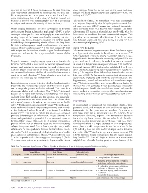
TABLE 6 Patterns and Significance of Tracer Uptake in Multiphase Bone Scintigraphy Applied in Frostbite Injury*
Tracer Phase
Blood Flow Soft Tissue Delayed (2–4 h
Category (1–60 sec postinjection) (3–10 min postinjection) postinjection) Physiologic Significance
1 Normal Normal Normal • Completely viable tissue
• Reactive hyperemia from reversible
2 Increased Increase Normal to mildly increased
soft-tissue ischemia; bone viable.
• If early after injury, deep soft-tissue
ischemia or hibernating tissue
3 Absent to diminished Absent to diminished Normal to mildly increased • If late after injury, deep soft-tissue
infarction
• Reversible bone ischemia
4 Absent Absent Absent Deep soft-tissue and bone damage
*Modified from Millet et al. 68
130 | JSOM Volume 20, Edition 4 / Winter 2020