
Page 74 - JSOM Fall 2020
P. 74
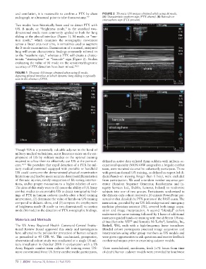
and ventilation, it is reasonable to confirm a PTX by chest   FIGURE 2  Thoracic U/S images obtained while using M mode.
          radiograph or ultrasound prior to tube thoracostomy.” 9  (A) Characteristic seashore sign (PTX absent). (B) Barcode or
                                                             stratosphere sign (PTX present).
          Two modes have historically been used to detect PTX with
          U/S. B mode, or “brightness mode,” is the standard two-   (A)
          dimensional mode most commonly applied to look for lung
          sliding at the pleural interface (Figure 1). M mode, or “mo-
          tion mode,” which examines the sonographic movement
          across a linear area over time, is sometimes used to augment
          the B-mode examination. Examination of a normal, uninjured
          lung will create characteristic findings commonly referred to
          as the “seashore sign,” whereas a PTX will create a charac-
          teristic “stratosphere” or “barcode” sign (Figure 2). Studies
          evaluating the value of M mode on the sensitivity/diagnostic
          accuracy of PTX detection have been mixed. 10,11
          FIGURE 1  Thoracic U/S image obtained when using B mode,
          depicting pleural interface at which dynamic lung sliding is typically
          seen in the absence of PTX.


                                                              (B)


          Though U/S is a potentially valuable adjunct in the hand of
          military medical technicians, scant literature exists on the em-
          ployment of U/S by military medics or the optimal training
          required to allow them to effectively use U/S at the point-of-  defined as active duty enlisted Army soldiers with military oc-
          care. 12,13  We postulate that rapid detection of a PTX by mil-  cupational specialty (MOS) 68W assigned to a brigade combat
          itary medical personnel equipped with portable or handheld   team, were recruited via email to voluntarily participate. Those
          U/S could overcome the demonstrated physical examination   with previous formal U/S training, as defined as expert-led di-
          limitations and lead to more accurate detection/differentiation   dactic/hands-on training longer than 1 hour, were excluded
          of thoracic injuries, timely integration of life-saving interven-  from participation. We used a random number sequence gen-
          tions, and/or proper evacuation to a higher echelon of care.   erator (Random Sequence Generator, Randomness and In-
          The aims of this study were to (1) assess the ability of US Army   tegrity Services Ltd., Dublin, Leinster, Ireland) to randomize
          combat medics to use portable U/S to detect sonographic find-  subjects into one of two groups. Participants randomized to
          ings of PTX in human cadaver models after a brief training   the didactic-only cohort received a 20-minute PowerPoint pre-
          intervention, (2) determine the value of hands-on U/S training   sentation that detailed the PTX portion of the FAST exam. The
          compared to didactic alone, and (3) compare the employment   instruction, provided by an U/S fellowship-trained emergency
          of brightness mode (B mode or two-dimensional) vs motion   medicine physician assistant (PA), covered both image acqui-
          mode (M mode) in the detection of PTX sonographic findings.  sition and image interpretation. A second “blended” cohort
                                                             underwent the same training followed by 1 hour of additional
          Materials and Methods                              instructor-guided hands-on training with two different U/S ma-
                                                             chines (Sonosite iVIZ  and Sonosite M-Turbo , SonoSite, Inc,
                                                                                                 ®
                                                                              ®
          The  US  Army  Regional  Health  Command-Central  Institu-  Bothell, WA), each with a high-frequency linear transducer.
          tional Review Board approved this study and investigators   Blended cohort participants practiced image acquisition and
          have adhered to the policies for protection of human subjects   interpretation using other group members as U/S models and
          as prescribed in 45 CFR 46. This randomized, prospective,   were given opportunities to ask questions and receive feedback
          observational cohort study was conducted at a single US mil-  on their techniques prior to examining cadaver models.
          itary installation in October 2018 in conjunction with a US
          Army Brigade combat team cadaver lab training event. U/S-   Three unembalmed, nonfrozen, fresh (<72 hours from time
          naive conventional force US Army combat medic participants,   of death) human cadaver models were provided by Southwest


          72  |  JSOM   Volume 20, Edition 3 / Fall 2020