
Page 82 - JSOM Spring 2018
P. 82
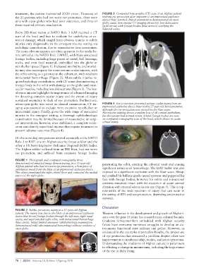
treatment, the patient maintained 20/20 vision. However, of   FIGURE 3  Computed tomography (CT) scan of an Afghan patient
          the 21 patients who had not worn eye protection, there were   without eye protection after exposure to an improvised explosive
          nine with open globes who had poor outcomes, and three of   device blast. Limited clinical examination demonstrated an open
          these required primary enucleation.                globe; axial, noncontrast CT imaging shows left lens dislocation
                                                             (gold arrow) with foreign bodies (blue arrows) overlying the
                                                             bilateral sclera.
          Every IED-blast victim at NATO Role 3 KAF received a CT
          scan of the head and face to evaluate for underlying or ex-
          tent of damage, which ranged from obvious trauma to subtle
          injuries only diagnosable in the emergent trauma setting via
          radiologic consultation, due to resuscitative time constraints.
          The more-obvious injuries are often apparent to the medic be-
          fore arrival at the NATO Role 3 MMU, with blast-associated
          foreign bodies, including large pieces of metal, ball bearings,
          rocks, and even fecal material, embedded into the globe or
          retrobulbar space (Figure 1). Increased morbidity and mortal-
          ity may also accompany the more serious ocular injuries, with
          the orbit serving as a gateway to the calvarium, with resultant
          intracranial hemorrhage (Figure 2). More-subtle injuries re-
          quired radiology consultation, with CT scans demonstrating a
          foreign body in the orbit with damage to the globe and extra-
          ocular muscles, including lens dislocations (Figure 3). The less-
          obvious injuries highlight the importance of advanced imaging
          for detecting complex ocular injury and the extent of injury
          sustained secondary to lack of eye protection. Furthermore,
          when open globe was noted on clinical examination, CT im-  FIGURE 4  Eye protection prevented serious ocular injury from an
          aging was essential to exclude more extensive retrobulbar or   improvised explosive device blast in this 27-year-old Servicemember,
          intracranial injury. Finally, given the wide range of associated   although the eye protection was ejected by the blast wave.
                                                             Fluorescein staining shows complete loss of all corneal epithelium,
          injuries in the emergent setting, a thorough ophthalmologic   but the patient had normal vision. Scleral foreign bodies are seen
          examination may be limited because of resuscitative or surgi-  on computed tomography scan of the head, which shows no acute
          cal interventions; however, once stabilized, a complete evalu-  orbital injury.
          ation can identify superficial injuries that require treatment to
          prevent adverse outcomes (Figure 4).

          On the same day, two patients arrived separately at the NATO
          Role 3 at KAF: one an Afghan security forces soldier and the
          other a US Navy Explosive Ordnance Disposal (EOD) Sailor.
          The Afghan soldier suffered from an IED blast, had not worn
          eye protection, and suffered from extensive foreign bodies

          FIGURE 1  Photograph and computed tomography three-
          dimensional reformatted image demonstrating, in a 35-year-old   penetrating the orbit, entering the calvarial vault and causing
          Afghan patient who had not worn eye protection, a 4cm piece of   significant intracranial hemorrhage. The EOD Sailor was also
          curvilinear metal from the blast of an improvised explosive device.
          The object penetrated the right orbital floor and contacted the medial   exposed to a significant explosion with the blast wave. Shrap-
          aspect of the right globe.                         nel crushed his ballistic-grade–issued eyewear and peppered his
                                                             face with foreign bodies; however, his orbits and intracranial
                                                             contents remained intact with the exception of acute corneal
                                                             abrasion with stromal edema in one eye (Figure 5). This is rep-
                                                             resentative  of  the  wide  spectrum  of  injury  that  can  occur  in
                                                             the setting of IED and eye protection, depending on protective
                                                             eyewear.

                                                             Discussion
          FIGURE 2  Subtle, yet severe, injury in a 27-year-old Afghan
          patient. The injury was due to the blast of an improvised explosive   Western influence in the development and growth of Afghani-
          device that forced foreign bodies through the left nare, right nasal   stan over the past 10 years has crossed many cultural bounds.
          cavity, and superomedial right orbit, with intracranial hemorrhage   Coalition Servicemembers  embedded with Afghan  security
          (blue circle). Initial limited emergent ophthalmology examination
          demonstrated mild subconjunctival hemorrhage without evidence of   forces have overcome numerous struggles to develop an au-
          open globe.                                        tonomous functional state military and police. However, as
                                                             evidenced in the examples reported in Results, the proper use
                                                             of eye protection has remained a problem despite other vast
                                                             improvements in marksmanship, tactics, and joint operations.
                                                             Understanding the traditions of Afghan culture is paramount
                                                             to effecting a change in mannerisms, including the importance
                                                             of the eye in daily living.


          78  |  JSOM   Volume 18, Edition 1/Spring 2018