
Page 85 - Journal of Special Operations Medicine - Fall 2016
P. 85
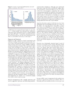
Figure 3  Location of exertional rhabdomyolysis cases and   and electrolyte imbalances. Although some individuals
               month of occurrence, 2011–2015. 35                 with lower serum CK levels (<10,000U/L), milder clini-
                                                                  cal presentations, and good follow-up may be treated
                                                                  in the clinic and managed as outpatients, the majority
                                                                  of presentations should be admitted to the hospital for
                                                                  inpatient care. A knowledgeable provider should be di-
                                                                  rectly involved in clinical care or available as a consul-
                                                                  tant; patients need to be observed carefully for clinical
                                                                  progress with special attention to screening for the sec-
                                                                  ondary development of compartment syndrome or iden-
                                                                  tifying individuals who may require dialysis.

                                                                  Aggressive hydration replaces fluids that may have been
                                                                  sequestered into the damaged myocytes (as a result of
                                                                  the failure of energy-dependent  transcellular pumps)
                                                                  and reduces the probability of acute kidney injury by
                                                                  increasing urine flow to assist in removal of myoglobin.
               and ER have been proposed, based largely on case stud-  To minimize the possibility of acute kidney injury, urine
               ies. 45,46  Civilian studies have indicated that individuals   output treatment goals should be >300mL/hr and urine
               with lower levels of physical activity or physical fitness   pH >7.5. The clinician should carefully direct therapy
               have higher ER risk. 12,29  A possible association between   guided by clinical response, including monitoring CK
               ER and sickle-cell trait has also been hypothesized, but   levels, renal function, and metabolic response. CK lev-
               there is considerable controversy on this topic. 47  els typically peak 2 to 3 days into the clinical presen-
                                                                  tation. A secondary rise in CK levels during treatment
               Diagnosis and Treatment                            should raise clinical suspicion of an occult compartment
               Diagnosis of ER is based on clinical examination and   syndrome.
               laboratory finding. Patients typically present with a his-
               tory of heavy and unaccustomed exercise and with symp-  Diuretics (e.g., furosemide, mannitol) may be used, if
               toms of severe muscle pain, muscle swelling, weakness,   necessary, to maintain urinary output. Mannitol, which
               and decreased range of motion. Their urine may be dark   increases renal blood flow and glomerular filtration
               brown (often described as “cola colored’). Pain is often   rate, is an osmotic agent that extracts fluids from in-
               localized to the muscle groups that were involved in the   terstitial compartments (thus reducing hypovolemia,
               heavy physical activity. Suggested diagnostic criteria for   muscle swelling, and nerve compression), and increases
               ER have been developed by the Uniformed Services Uni-  urinary flow (reducing myoglobin precipitation). Urine
               versity Consortium for Health and Military Performance   can be alkalinized by the addition of 50–100mEq of
               (CHAMP) in conjunction with the Israeli Defense Force’s   sodium bicarbonate to each liter of administered fluid.
               Heller Institute. These criteria include the requisite clini-  The proposed ideal fluid regimen is half isotonic saline
               cal presentation in conjunction with (1) a serum CK level   (0.45% sodium), to which 75mmol/L sodium bicar-
               5 times higher than the upper limit of normal and/or (2)   bonate is added. Although mannitol and bicarbonates
               a urine dipstick positive for blood (due to the presence   are the standard of care for reducing the likelihood of
               of myoglobin) but lacking red blood cells under micro-  acute renal failure, some studies suggest that their use
               scopic urinalysis.  Two distinct subgroups of ER have   provides no additional benefit to patients over aggres-
                              48
               been suggested by the CHAMP and Heller Institute: (1)   sive hydration with  saline alone. Blood urea  nitrogen
               physiologic (benign) ER and (2) clinically relevant ER.   and creatinine levels can be monitored to indicate re-
               Physiological ER is defined as a patient with an elevated   nal function. Attention should be directed to monitor-
               CK level but no other signs or symptoms beyond mild   ing potassium, calcium, and phosphate levels to correct
               muscle pain expected for the circumstances. This is es-  hyperkalemia, hypocalcemia, and hypophosphatemia
               sentially delayed-onset muscle soreness. In clinically rel-  when  present.  Hyperkalemia  and hypophosphatemia
               evant ER, the patient presents with severe muscle pain,   result from direct release of potassium and phosphates
               muscle swelling, muscle weakness, and myoglobinuria,   from muscles. Hypocalcemia results from the buildup of
               along with the other diagnostic criteria.  It is likely that   calcium in muscle due to the failure of sodium-calcium
                                                 48
               these distinctions are “points” on a continuum ranging   exchange. 1–3,14,49–52
               from mild to severe muscle damage.
                                                                  In cases of ER, it may be important for the medical care
               Medical management  of ER is largely supportive  and   provider to question the patient as to whether other in-
               involves aggressive hydration and addressing metabolic   dividuals performed similar activities or are using a new


               Exertional Rhabdomyolysis                                                                        67