
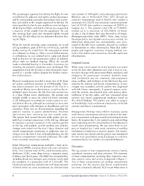
Page 24 - Journal of Special Operations Medicine - Fall 2016
P. 24
The paratrooper reported that during the flight, he was   total protein of 100mg/dL, and a decreased glomerular
                                                                                              2
            immobilized by adjacent and tightly packed passengers   filtration rate of 64mL/min/1.73m . LFTs showed an
            and by surrounding rucksacks that pinned him in posi-  aspartate transaminase level of 862U/L that reached a
            tion and added to the weight supported by his lap. He   maximum of 1,762U/L and an alanine aminotransferase
            noted specifically that he was unable to move his legs at   level of 233U/L that reached a maximum of 520U/L on
            all during the flight, and that his left leg was supporting   hospital day 2. The patient’s total CPK of 77,900U/L
            a majority of the weight from the equipment. He was   trended up to a maximum of 102,380U/L on hospi-
            also wearing knee pads that wrapped tightly around   tal day 2. His D-dimer level was elevated to 19.6mg/L
            both legs. He fell asleep for an unknown duration dur-  fibrinogen-equivalent units (FEU). Urine drug screen,
            ing the flight.                                    blood gas levels, and hepatitis screens were normal. An
                                                               abdominal ultrasound was unremarkable, and ultra-
            When he started receiving jump commands, he noted   sound of the left lower extremity showed no evidence
            left leg numbness, pain of 8–9 on a 0–10 scale, and dif-  of thrombosis or other abnormality. Plain film radio-
            ficulty moving the distal limb. He attempted to stand   graphs and magnetic resonance imaging without con-
            but collapsed on doing so. After a second failed attempt   trast of the lumbar spine showed no clinically significant
            at ambulation, he was ordered not to jump and placed   abnormalities.
            back in his seat by the jumpmaster safety, an infantry
            officer with no medical training. When the aircraft   Hospital Course
            made a scheduled landing in Alexandria, Louisiana, for
            refueling, the patient’s symptoms were unchanged. The   While some improvement in motor function was noted
            jumpmaster made the decision to have the patient trans-  in the left foot after admission and after 2 days of con-
            ported to a nearby civilian hospital for further evalua-  servative therapy with intravenous fluids, mannitol, and
            tion and treatment.                                analgesics, the paratrooper remained markedly weak
                                                               with  dorsiflexion  and  toe  extension,  and  progressive
            Physical examination revealed a heart rate of 96 beats   pain, swelling, and erythema of the left lower leg were
            per minute and blood pressure of 139/80mmHg. There   noted in the anterior and lateral compartments. A neu-
            was absent sensation over the dorsum of the foot in a   rologist was consulted and the patient was diagnosed
            superficial fibular nerve distribution, as well as the in-  with left fibular neuropathy. A general surgeon evalu-
            terdigital space between the left first and second toes   ated the patient, documented pain with passive plan-
            in a deep fibular nerve distribution. The patient was   tarflexion of the left ankle, and was concerned about
            initially unable to move the distal left lower extremity   anterior  and  lateral  compartment  syndrome  based on
            at all. Subsequent re-evaluations noted a return to nor-  the clinical findings. The patient was taken to surgery
            mal plantar flexion, although he continued to have only   on hospital day 3 and underwent a fasciotomy of the left
            trace movement with attempts at dorsiflexion and toe   anterior and lateral compartments.
            extension. There was no documentation regarding the
            patient’s response to passive plantarflexion. Inversion   Intraoperative findings noted mild to moderate edema
            and eversion  were improving but also  notably weak.   in the lateral compartment and severe edema in the ante-
            The patient had normal dorsalis pedis pulses and ini-  rior compartment with gray muscle protruding from the
            tially had a normal appearance of the left leg, although   fascia. By hospital day 5, the patient’s pain and swelling
            he reported a pressure sensation in the anterior portion   were improving and the abnormalities in his laboratory
            of the leg, and gradual, painful swelling and cellulitic   results were downtrending. He was taken back to sur-
            changes were noted over the next 2 days. Anterior and   gery on hospital day 5 with noted reduction of swelling
            lateral compartment tenderness to palpation also de-  and absence of infection or necrotic muscle. The fasciot-
            veloped over the first 2 days of hospitalization, but the   omy wound was closed and the patient was monitored
            posterior compartment remained unaffected. The right   for two more days before being discharged to a military
            lower extremity functioned normally.               treatment facility for further care and rehabilitation.

            Initial laboratory examinations included a basic meta-  Discussion
            bolic panel (BMP), complete blood cell count, urinalysis
            (UA), liver function tests (LFTs), total keratinize phos-  The leg consists of four compartments: anterior, lateral,
            phokinase (CPK), urine drug screen, a hepatitis panel,   superficial posterior, and deep posterior. The anatomy
            D-dimer, and blood gases. The BMP was unremarkable,   of these compartments, including their respective mus-
            including blood urea nitrogen and creatinine levels, with   cles, arteries, veins, and nerves, is depicted in Figure 1.
            the exception of a potassium level of 5.3mmol/L. The   Any of these compartments can develop compartment
            UA showed grossly tea-colored urine with large amounts   syndrome, but the anterior and lateral compartments
            of blood noted on urine microscopy, an  elevated urine   are more commonly affected. ACS occurs when there is


            6                                         Journal of Special Operations Medicine  Volume 15, Edition 3/Fall 2016