
Page 20 - Journal of Special Operations Medicine - Fall 2016
P. 20
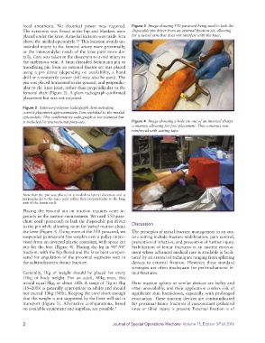
local anesthesia. No electrical power was required.   Figure 3  Image showing 550 paracord being used to lash the
            The extremity was flexed at the hip and blankets were   disposable pin driver from an external fixation set, allowing
            placed under the knee. A medial incision was made 3cm   for a swivel arm that does not interfere with the knee.
                                     3,4
            above the medial epicondyle.  This location avoids un-
            intended injury to the femoral artery more proximally,
            or the intercondylar notch of the knee joint more dis-
            tally. Care was taken in the dissection to avoid injury to
            the saphenous vein. A 5mm threaded Steinmann pin or
            transfixing pin from an external fixator set was placed
            using a pin driver (depending on availability, a hand
            drill or a nonsterile power drill may also be used). The
            pin was placed horizontal to the ground, and perpendic-
            ular to the knee joint, rather than perpendicular to the
            femoral shaft (Figure 2). A plain radiograph confirmed
            placement but was not required.

            Figure 2  Anterior-posterior radiograph demonstrating
            correct placement approximately 3cm cephalad to the medial
            epicondyle. This confirmatory radiograph is not required but
            is included for instructional purposes.            Figure 4  Image showing a hole cut out of an inverted sharps
                                                               container, allowing for foot placement. This construct was
                                                               reinforced with casting tape.


            Note that the pin was placed in a medial to lateral direction and is
            perpendicular to the knee joint rather than perpendicular to the long
            axis of the femur itself.

            Placing the femoral pin on traction requires some in-
            genuity in the austere environment. We used 550 para-
            chute cord (paracord) to lash the disposable pin driver   Discussion
            to the pin while allowing room for swivel motion about
            the knee (Figure 3). Using more of the 550 paracord, we   The principles of initial fracture management in an aus-
            suspended gymnasium free weights over a pulley impro-  tere setting include fracture stabilization, pain control,
            vised from an inverted plastic container, with space cut   prevention of infection, and prevention of further injury.
            out for the foot (Figure 4). Placing the leg in 90°-90°   Stabilization of femur fractures in an austere environ-
            traction, with the hip flexed and the knee bent compen-  ment where advanced medical care is available is facili-
            sated for angulation of the proximal segments seen in   tated by an arsenal of techniques ranging from splinting
            the subtrochanteric femur fracture.                devices to external fixation. However, these standard
                                                               strategies are often inadequate for peritrochanteric fe-
            Generally, 1kg of weight should be placed for every   mur fractures.
            10kg of body weight. For an adult, 80kg man, this
            would equal 8kg, or about 18lb. A range of 7kg to 9kg   Hare traction splints or similar devices are bulky and
            (15–20lb) is generally appropriate in adults and should   often unavailable, and their application confers risk of
            not exceed 13kg (30lb). Keeping the cord short enough   significant skin breakdown, especially with prolonged
            that the weight is not supported by the litter will aid in   evacuation. These traction devices are contraindicated
            transport (Figure 5). Alternative configurations, based   for proximal femur fractures if concomitant ipsilateral
            on available equipment and supplies, are possible. 5  knee or tibial injury is present. External fixation is of


            2                                         Journal of Special Operations Medicine  Volume 15, Edition 3/Fall 2016